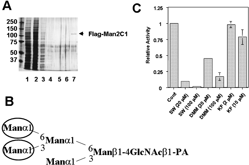
Figure 2. Purification and characterization of FLAG–Man2C1.
(A) Silver staining pattern of purified FLAG–Man2C1. Samples, 5 μl (lane 1) or 10 μl (lanes 2–7) were analysed using 7.5% PAGE. Lane M: molecular mass marker; lane 1, cell extract; lane 2, cytosolic fraction; lanes 3–6, first–fourth wash fractions of anti-FLAG antibody beads; lane 7, eluted fraction. (B) Structure of Man5GlcNAc-PA prepared from RNase B. Man residues with circles are reported to be cleaved by cytoplasmic α-mannosidase only in the presence of Co2+. (C) Effect of various mannosidase inhibitors on the activity of purified FLAG–Man2C1.

