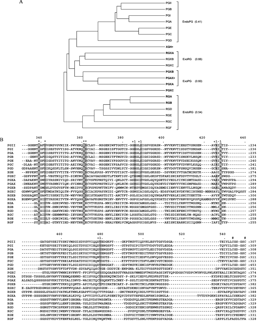
Figure 1. Comparative sequence analysis of A. niger family 28 glycoside hydrolases.
(A) Dendrogram of A. niger family 28 glycoside hydrolases. The average pairwise distance within the different functional groups of A. niger hydrolases is indicated in brackets. Biochemically identified enzymes are in bold. *A. niger enzyme amino acid sequences that are greater than or equal to 97% identical to that of a previously characterized A. tubingensis enzyme. (B) Region of the multiple alignment of A. niger family 28 glycoside hydrolases. *Columns containing catalytic residues; #conserved cysteine bridge; +1, columns containing residues involved in substrate binding at subsite +1; −1, columns containing residues involved in substrate binding at subsite −1. The complete multiple alignment of A. niger family 28 glycoside hydrolases is provided as Supplementary Figure 1 (http://www.BiochemJ.org/bj/400/bj4000043add.htm).

