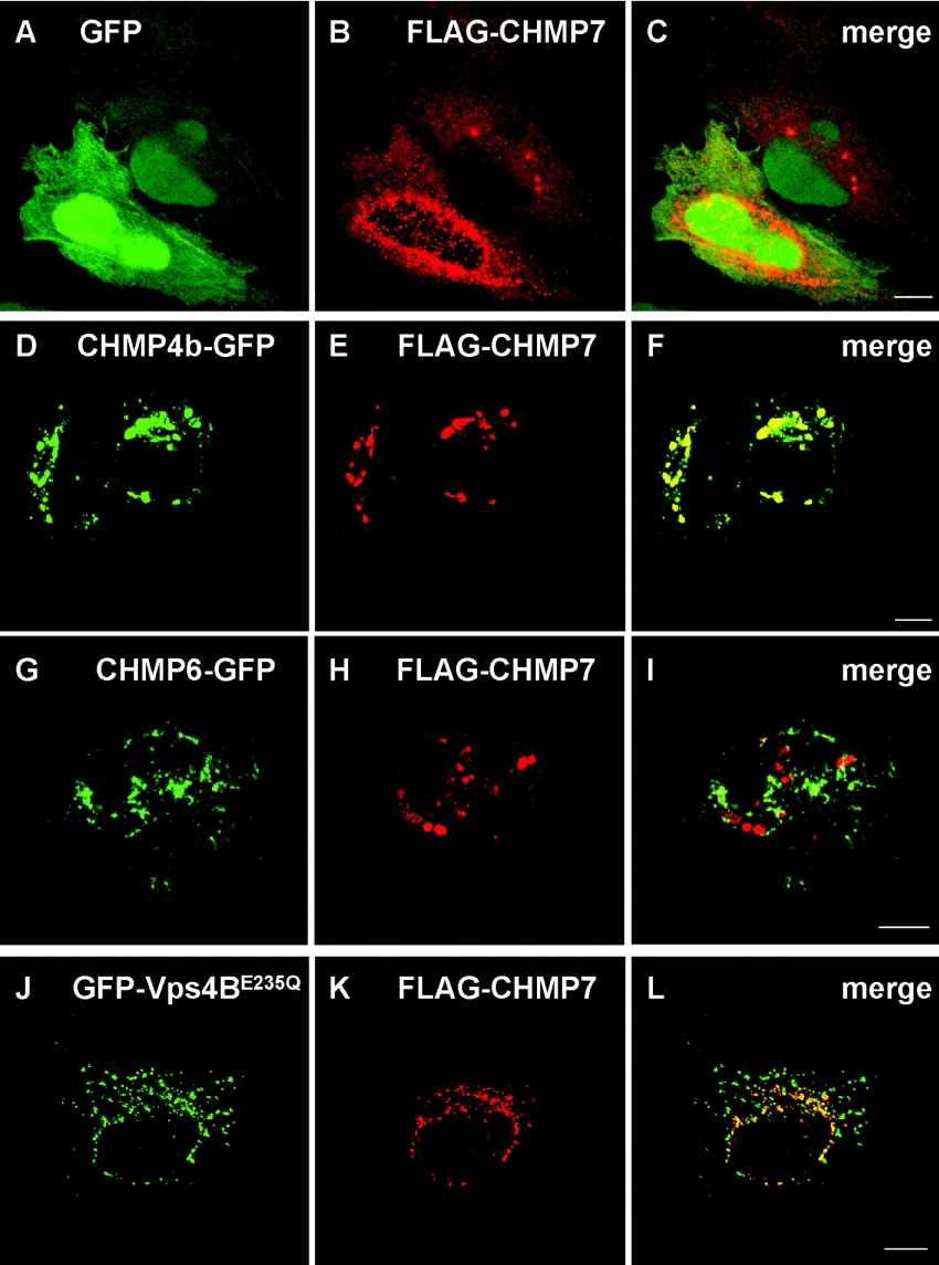
Figure 3. Subcellular localization of CHMP7.
HeLa cells were co-transfected with plasmids encoding FLAG–CHMP7 and GFP (A–C), CHMP4b–GFP (D–F), CHMP6–GFP (G–I) or GFP–Vps4BE235Q (J–L). After 24 h, the cells were fixed and subjected to immunofluorescence confocal microscopic analysis by direct visualization of GFP or GFP-fusion proteins (A, D, G and J; green) and by staining with anti-FLAG mAb (M2) and Cy3-labelled goat anti-mouse IgG antibody (B, E, H and K; red). Their merged images are shown in (C, F, I and L) respectively. Scale bars, 10 μm.

