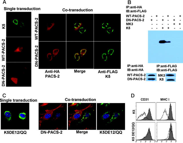
Figure 7.
PACS-2 interacts with acidic clusters in the carboxyterminus of K5. E-DMVECs were transduced with the indicated recombinant Ad constructs either alone (left panel) or in combination (right panel). After 24 hours of infection, cells were permeabilized and K5 was detected using anti-FLAG (green), whereas PACS-2 was visualized using anti-HA (red). Note that DN-PACS-2 localization changes from cytoplasmic to perinuclear in cells infected with AdK5. Merging the images (center) indicates colocalization of DN-PACS-2, but not WT-PACS-2, with K5. (B) CD31 was immunoprecipitated using anti-HA from E-DMVECs transduced with the indicated adenovirus constructs. Coprecipitated K5 was detected using anti-FLAG by immunoblotting the PACS-2 preciptiates. In contrast, MK3 was absent. Control blots using anti-FLAG and anti-HA antibodies confirm expression of the respective proteins. (C) DN-PACS-2 (red) does not colocalize with K5DE12/QQ (green). (D) CD31 and MHC I expression at the cell surface was monitored by flow cytometry at 24 hours after transduction of E-DMVECs with AdK5 or AdK5DE12 (unshaded). AdTet-infected cells were used as positive control (shaded).

