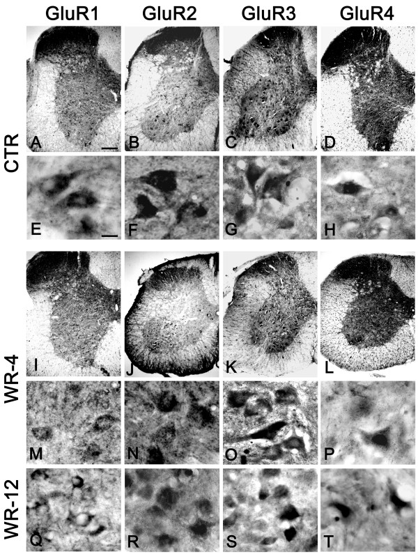
Figure 1.
AMPA receptor subunits in the spinal cord of control and wobbler mice. Representative photomicrographs showing the pattern of AMPA immunostaining in the cervical spinal cord of four-week-old healthy mice (A-D) and age-matched wobbler mice (I-L). Subcellular localisation of the different AMPA receptor subunits in four-week-old healthy mice is shown in panels (E-H) Panels (M-P) show the pattern of staining in motoneurons of the cervical spinal cord from four-week-old wobbler mice. The distribution of GluR 1–4 in the cervical spinal cord of 12-week-old wobbler mice is shown in panels (Q, R, S, T). Scale bar, A-D, I-L 100 μm. E-H, M-T 20 μm.

