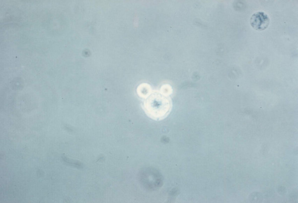
Figure 4.
LAK cell binding of MDA-MB-231 breast carcinoma cells. 1 × 10⁵ LAK cells (0.1 ml) and 1 × 10⁵ target cells (0.1 ml) were mixed in the same tube, centrifuged for 2 min at 500 g and incubated at 37°C for 10 min. The cells stained with trypan blue were observed under phase contrast microscope. 2 LAK cells forming a conjugate with a live tumor cell is demonstrated.

