
Figure 1.
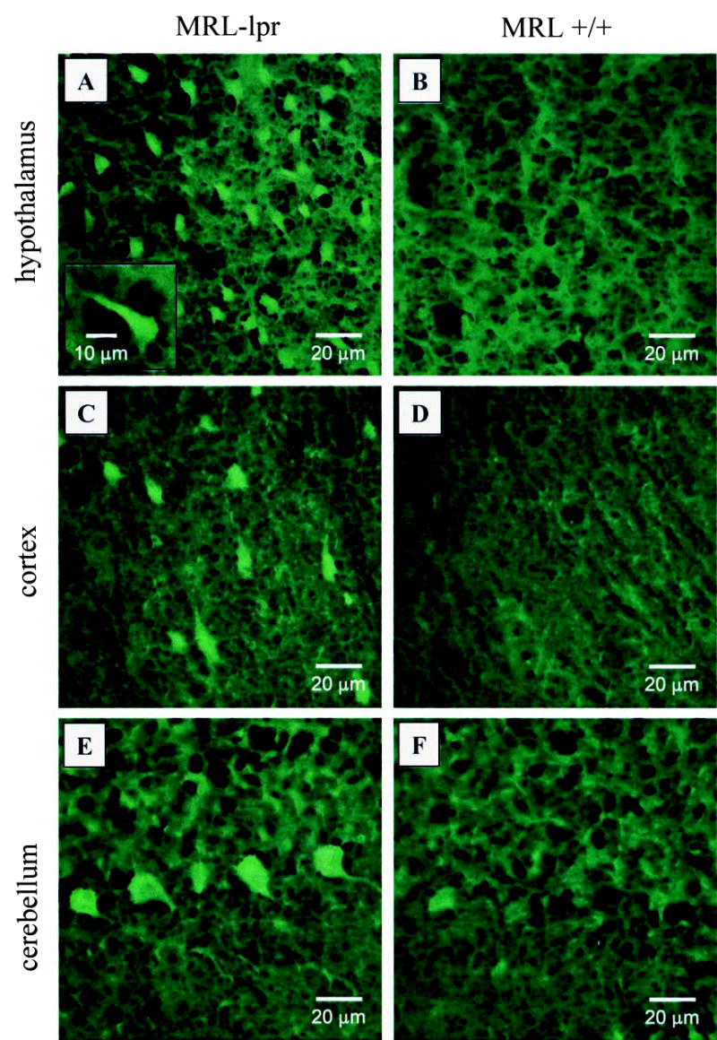
Representative fields of Fluoro-Jade B (FJB) staining in MRL brains. More numerous, brightly stained FJB+ cells suggest a neurodegenerative process in the diencephalon of MRL-lpr mice (A) in comparison to age and sex-matched MRL +/+ controls (B). Similarly, scattered FJB+ cells were commonly seen in the cortical parenchyma (C) in comparison to comparable areas from control brains (D). Some contiguous Purkinje cells were distinctly stained in MRL-lpr mice (E), but were not seen in congenic MRL +/+ controls (D).