
Figure 5.
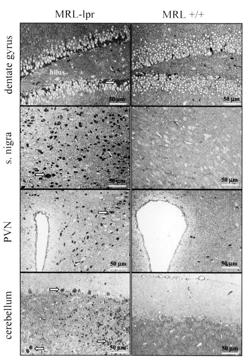
Toluidine blue staining of various brain regions inspected by light microscopy. The CA2/CA3 region (not shown) and subgranular zone of the dentate gyrus of 2 diseased MRL-lpr mice were frequently populated with densely packed, elongated dark cells. Although round, dark cells were clustered in the substantia nigra, they were scattered in the paraventricular nucleus (PVN), and Purkinje and granule cell layers of the cerebellum.