
Figure 7.
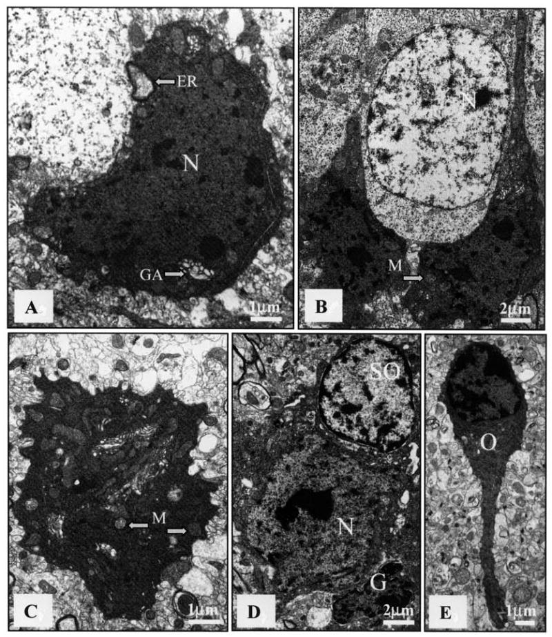
Electron microscopy (EM) revealing ultrastructural features of dark cells in brain of an MRL-lpr mouse. The increased electron density was unaccompanied by blebbing of cell membranes or internal changes in organelles, other than occasional swelling. A. Hypothalamic neuron (N) with densely compacted karyoplasm and cytoplasm, as well as an enlarged Golgi apparatus (GA) and endoplasmic reticulum (ER) (bar = 1 μm). B. Densely packed dark cells between healthy neurons (N) in the subgranular zone (bar = 2 μm). C. Cerebellar neuron with condensed cytoplasm, swollen mitochondria (M), and ruffled outer membrane (bar = 1 μm). D. Dark neuron (N) surrounded by healthy-looking satellite oligodendrocyte (SO) and shrunken glial cell (G) (bar = 2 μm). E. Hippocampal oligodendrocyte (O) with electron-dense cytoplasm and karyoplasm. No evidence of apoptotic bodies, nuclear fragmentation, or ruptured membranes could be observed in any preparation (bar = 1 μm).