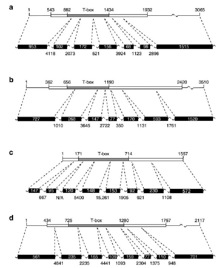
Fig. 1.

Genomic locus structure of Tbx1, Tbx2, Tbx5, and Tbx20 in Xenopus tropicalis. Tbx1 (a), Tbx2 (b), Tbx5 (c), and Tbx20 (d) cDNAs and their corresponding genomic loci are shown in diagrammatic form (not to scale). Coding regions of each cDNA are shown (boxes) together with their nucleotide positions and the position of the T-box (defined by alignment of the encoded proteins with the T-domain of Xbra) is also indicated. The exons corresponding to the cDNA sequences are shown together with their sizes (in base pairs) plus those of the intervening introns. Note that, as the size of the first exon of each gene is predicted based on the available cDNA sequence, the sizes of these exons may be underestimated here.
