
Figure 1. Melanocytes cultured from patients with Hermansky–Pudlak Syndrome (HPS)-1, HPS-2, and HPS-3 exhibit specific alterations in the distribution of tyrosinase gene family members.
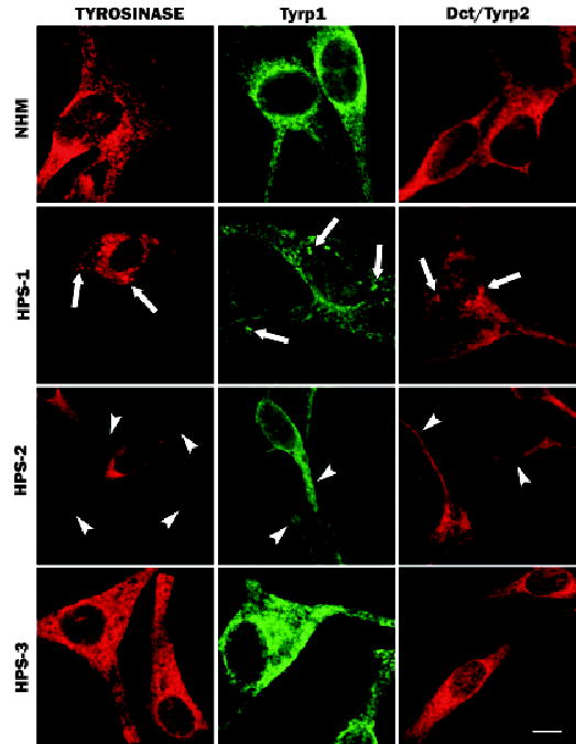
Cultures of melanocytes derived from an unaffected individual (normal human melanocytes (NHM)) and from patients with HPS-1, HPS-2, or HPS-3 were immunostained for tyrosinase, tyrosinase-related protein-1 (Tyrp1), or DOPAchrome tautomerase/tyrosinase-related protein-2 (Dct/Tyrp2). The NHM exhibited a granular staining pattern with prominent perinuclear localization for all three proteins. HPS-1 melanocytes exhibited a similar granular staining pattern with additional large aggregates (arrows) for all three proteins throughout the melanocytes. HPS-2 melanocytes exhibited a relative restriction of tyrosinase to the cell body with limited or no expression in the dendrites (arrowheads); Tyrp1 and Dct/Tyrp2 exhibited normal expression patterns. HPS-3 melanocytes displayed a floccular staining pattern within the cell body and dendrites for tyrosinase, Tyrp1, and Dct/Tyrp2 expression. Scale bar =10 μm.