
Figure 2. Hermansky–Pudlak Syndrome (HPS)-1 melanocytes exhibit a variation in co-localization of tyrosinase and tyrosinase-related protein-1 (Tyrp1) within characteristic large granules/membranous complexes.
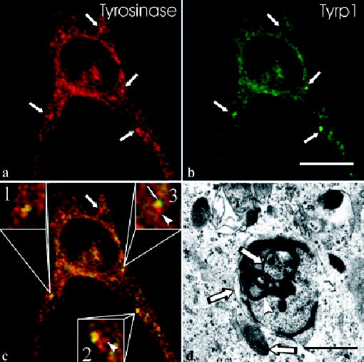
In HPS-1 melanocytes, the characteristic large granules (arrows) are positive for tyrosinase [red in (a)] and Tyrp1 [green in (b)]. The merged image (c) demonstrates that some large granules contained tyrosinase only (arrow), some show equal co-localization of tyrosinase and Tyrp1 (inset #1), some display prominent co-localization of tyrosinase and Tyrp1 with areas containing tyrosinase only (arrowhead, inset #2), and some show polar expression of tyrosinase (arrowhead, inset #3) and Tyrp1 (half-arrow) with an intervening area of co-localization. Electron microscopic evaluation of HPS-1 melanocytes treated for DOPA histochemistry (d) demonstrates that, within a membranous complex, some cisternae and melanosomes express (arrowheads) or lack (arrows) reaction product, indicating the presence and absence of tyrosinase, respectively. Scale bars =10 (b) and 1.5 (d) μm.