
Figure 3. Melanocytes cultured from patients with Hermansky–Pudlak Syndrome (HPS)-1, HPS-2, and HPS-3 exhibit specific alterations in the distribution of HPS gene products.
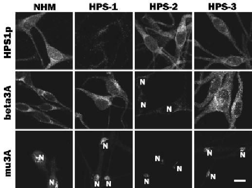
Cultures of melanocytes derived from an unaffected individual (normal human melanocytes (NHM)) and patients with HPS-1, HPS-2, or HPS-3 were immunostained for HPS1 (HPS-p) and the β3A (beta3A) and μ3A (mu3A) subunits of AP3. Expression of HPS1 protein (HPS1p) was similar in NHM, HPS-2 melanocytes, and HPS-3 melanocytes but markedly diminished in HPS-1 melanocytes. Expressions of both β3A and μ3A were normal in NHM, HPS-1 melanocytes, and HPS-3 melanocytes but markedly diminished in HPS-2 melanocytes. N, nucleus. Scale bar =10 μm.