
Figure 5. Melanocytes cultured from patients with Hermansky–Pudlak Syndrome (HPS)-1, HPS-2 and HPS-3 were evaluated for expression of various proteins.
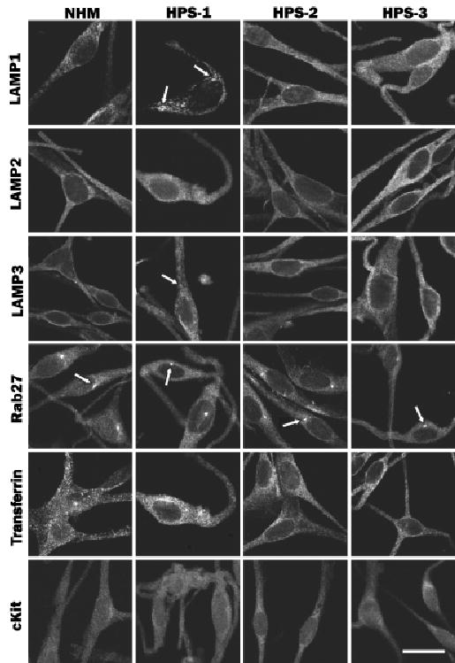
Normal human melanocytes (NHM) and HPS-1, HPS-2, or HPS-3 melanocytes were immunostained for LAMP 1–3, Rab 27, transferrin, and cKit. Expression of LAMP1 and LAMP3 was granular and that of LAMP2 was diffuse throughout the NHM. HPS-1 melanocytes exhibited, in addition to the normal localization for LAMP 1–3, localization to large granules (arrows) for LAMP1 and LAMP3. HPS-2 melanocytes exhibited normal localization for LAMP 1–3. HPS-3 melanocytes exhibited a more floccular distribution pattern for LAMP1 and LAMP3 and a normal distribution of LAMP2. Expression of Rab27, transferrin, and cKit in NHM exhibited a uniform pattern throughout the melanocytes with a distinct centriole localization (arrows), a punctate pattern throughout the melanocytes, and a diffuse pattern throughout the melanocytes, respectively. HPS-1, HPS-2, and HPS-3 melanocytes exhibit a normal staining pattern for Rab27, transferrin, and cKit. Scale bar =20 μm.