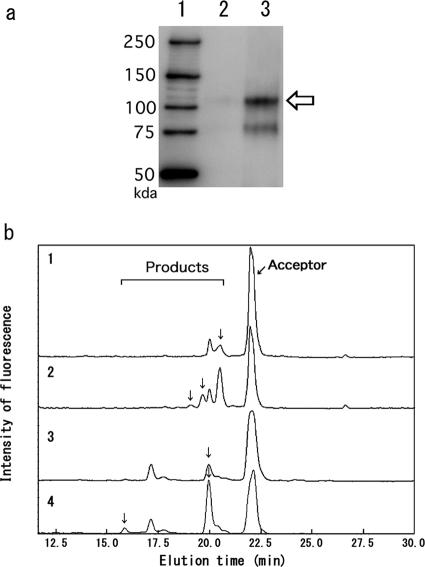
FIG. 2.
Effect of PDI on amount of Pir fusion protein in the cell wall. (a) An HA-tagged fusion protein (HA-pp-GalNAc-T10) was detected by Western blotting from YSF124 cells (lane 2) and from YSF124 cells with PDI overexpression (lane 3). Lane 1, molecular markers. Arrow indicates the fusion protein. (b) The enzymatic activities of pp-GalNAc-T6 and -T10 were analyzed by HPLC as described in Materials and Methods. Enzymatic activities were expressed as arbitrary units (1 arbitrary unit = 1 pmol of product/5 OD600 units of cell wall/24 h). Arrows indicate the peaks for acceptor and product oligosaccharides. Activities were as follows: for pp-GalNAc-T6 expressed in YSF123 cells (curve 1), 35 U; for pp-GalNAc-T6 expressed in YSF123 cells with PDI overexpression (curve 2), 150 U; for pp-GalNAc-T10 expressed in YSF124 cells (curve 3), 90 U; for pp- GalNAc-T10 expressed in YSF124 cells with PDI overexpression (curve 4), 230 U.

