Abstract
The outermost layer of the Bacillus anthracis spore is the exosporium, which is composed of a paracrystalline basal layer and an external hair-like nap. The filaments of the nap are formed by a collagen-like glycoprotein called BclA, while the basal layer contains several different proteins. One of the putative basal layer proteins is ExsY. In this study, we constructed a ΔexsY mutant of B. anthracis, which is devoid of ExsY, and examined the assembly of the exosporium on spores produced by this strain. Our results show that exosporium assembly on ΔexsY spores is aberrant, with assembly arrested after the formation of a cap-like fragment that covers one end of the forespore—always the end near the middle of the mother cell. The cap contains a normal hair-like nap but an irregular basal layer. The cap is retained on spores prepared on solid medium, even after spore purification, but it is lost from spores prepared in liquid medium. Microscopic inspection of ΔexsY spores prepared on solid medium revealed a fragile sac-like sublayer of the exosporium basal layer, to which caps were attached. Examination of purified ΔexsY spores devoid of exosporium showed that they lacked detectable levels of BclA and the basal layer proteins BxpB, BxpC, CotY, and inosine-uridine-preferring nucleoside hydrolase; however, these spores retained half the amount of alanine racemase presumed to be associated with the exosporium of wild-type spores. The ΔexsY mutation did not affect spore production and germination efficiencies or spore resistance but did influence the course of spore outgrowth.
Bacillus anthracis, the causative agent of anthrax, is a gram-positive, rod-shaped, aerobic bacterium that forms endospores (or spores) when vegetative cells are deprived of certain nutrients (22). Spore formation begins with an asymmetric septation in the starved cell that produces large and small genome-containing compartments, called the mother cell and forespore, respectively (31). The mother cell then engulfs the forespore and surrounds it with three concurrently synthesized layers, called the cortex, coat, and exosporium (5). The cortex, which is the innermost and thickest of the three layers, is composed of peptidoglycan (4). The coat, which tightly covers the cortex, is composed of an undetermined but probably large number of different proteins (14). The exosporium, which is a loose-fitting, balloon-like structure enclosing the spore, is apparently composed of at least a dozen different proteins and glycoproteins (29). After spore formation is complete, the mother cell lyses to release the spore. Mature spores are dormant and resistant to harsh chemicals and physical damage, which allows them to survive in their normal soil environment for many years (18). When spores encounter an aqueous environment containing appropriate nutrients, they germinate and grow as vegetative cells (27). Germination is activated by small-molecule germinants, such as l-alanine and inosine, which are recognized by receptors located within the spore membrane that underlies the cortex (36). When B. anthracis spores enter a human or other mammalian host, germination and cell growth produce toxins that can rapidly cause death (17).
Recent concerns about the use of B. anthracis spores as a biological weapon have resulted in efforts to better understand the interactions between B. anthracis spores and the cells of the mammalian immune system (2, 9, 12, 23) and also to develop better detectors for these spores (25, 30, 32, 37). Both efforts require a detailed molecular description of the outermost layer of the B. anthracis spore, the exosporium. The exosporium serves as a semipermeable barrier that excludes large, potentially harmful molecules, such as antibodies and hydrolytic enzymes (5, 6), and it also serves as the source of surface antigens (5, 28, 32). The exosporium is composed of a paracrystalline basal layer and an external hair-like nap. Most, if not all, of the filaments of the hair-like nap are formed by a single collagen-like glycoprotein called BclA (1, 33). In contrast, the basal layer appears to be composed of at least a dozen different proteins in tight and loose associations (24, 29). The proteins include BxpB (also called ExsF), which was recently shown to be required for the attachment of BclA and the hair-like nap to the basal layer and also to play a role in suppressing spore germination (29, 34). In the exosporium, BclA and BxpB are present in stable high-molecular-mass (i.e., >250 kDa) complexes, which also contain the protein ExsY and, possibly, its homologue CotY (24, 29). It has been reported that ExsY is required for exosporium assembly in Bacillus cereus, which forms spores very similar to those of B. anthracis (35).
In this study, we constructed a ΔexsY mutant strain of B. anthracis and used this strain to examine the role of ExsY in exosporium assembly. Our results show that in sporulating cells devoid of ExsY, exosporium assembly is arrested after the formation of a cap-like fragment that covers one end of the forespore. Inspection of the resulting spores revealed new structural features within the exosporium basal layer. The ΔexsY spores could readily be stripped of apparently all exosporium. These exosporiumless spores were compared to wild-type spores to identify exosporium proteins and functions. Other characteristics of ΔexsY spores were examined, including variations in the outgrowth of germinated spores. A possible mechanism for the assembly of the exosporium is discussed.
MATERIALS AND METHODS
B. anthracis strains.
The Sterne 34F2 veterinary vaccine strain of B. anthracis was obtained from the U.S. Army Medical Research Institute of Infectious Diseases, Fort Detrick, Md., and used as the wild-type strain in this study. The Sterne strain is not a human pathogen because it lacks plasmid pXO2, which is necessary to produce the capsule of the vegetative cell (8). A variant of the Sterne strain carrying a ΔexsY mutation that precisely deletes the entire exsY open reading frame was constructed by allelic replacement, essentially as previously described (3, 29). This procedure replaced the exsY gene with a spectinomycin resistance cassette, which was confirmed by PCR amplification and DNA sequencing of the relevant region of the chromosome. The ΔexsY mutant strain was called CLT325.
Preparation of spores and sporulating cells.
Spores were prepared by growing strains at 37°C in liquid (with shaking) or on solid (1.5% agar) Difco sporulation medium (DSM) (19) until sporulation was complete, typically 48 to 72 h. Spores were collected by centrifugation, washed extensively with cold (4°C) sterile deionized water, sedimented through a two-step gradient of 20% and 50% Renografin (Bracco Diagnostics), and extensively washed again with cold water (10). Spores were stored and quantitated as previously described (28). Sporulating cells, also called sporangia, were obtained from cultures grown in liquid DSM at 37°C with shaking. Culture density was measured spectrophotometrically at 600 nm, and spore development was monitored by phase-contrast microscopy. Sporangia were harvested by centrifugation at 4,000 × g for 10 min at 4°C.
Preparation of recombinant exosporium proteins.
Recombinant versions of the B. anthracis exosporium proteins ExsY, CotY, and alanine racemase (Alr) were prepared essentially as previously described (28, 29). Briefly, the genes encoding these proteins (i.e., BAS1141, BAS1145, and BAS238, respectively [B. anthracis Sterne gene numbers from the Kyoto Encyclopedia of Genes and Genomes database {11}]) were amplified by PCR and inserted into the cloning site of the expression vector pET15b (Novagen). The resulting plasmids were transformed individually into Escherichia coli strain BL21(DE3) to express the cloned genes according to the pET system manual (Novagen). Each expressed recombinant protein contained a six-His tag and a factor Xa cleavage site immediately preceding the initiating methionine. To isolate recombinant ExsY (rExsY) and rCotY, cells expressing individual proteins were broken by sonication, and insoluble cellular material, which contained essentially all of the recombinant protein, was recovered by centrifugation at 10,000 × g for 20 min at 4°C. The insoluble material, the vast majority of which was the recombinant protein, was resuspended in 8 M urea and dialyzed against phosphate-buffered saline (PBS) (26) prior to use. Recombinant Alr was purified under native conditions by immobilized-metal affinity chromatography (QIAGEN), and its six-His tag was removed by factor Xa cleavage as previously described (29).
Preparation of mouse MAbs.
The production and characterization of anti-BclA and anti-BxpB monoclonal antibodies (MAbs), designated EF12 and 10-23-4, respectively, were described previously (1, 28, 29). MAb 10-23-4 does not react with a paralogue of BxpB that is encoded by the BAS2303 gene and is 78% identical to BxpB (29). An anti-BxpC MAb designated FH6-1 was raised against purified Sterne exosporium by using a published procedure (28). FH6-1 was shown to bind the B. anthracis BxpC protein and a recombinant version synthesized in E. coli (C. T. Steichen and C. L. Turnbough, Jr., unpublished data). Purified rExsY and rAlr were used as antigens to raise an anti-ExsY/CotY MAb, designated G9-3, and an anti-Alr MAb, called AR-1, according to published procedures (29). In immunoblots, G9-3 reacted similarly with rExsY and rCotY, which are 85% identical; AR-1 did not react with a second B. anthracis alanine racemase that is encoded by the BAS1932 gene and apparently produced during vegetative growth. All MAbs were purified by affinity chromatography on protein G-Sepharose (28, 32). The MAbs represented three isotypes, i.e., immunoglobulin G1 (IgG1), κ chain (10-23-4, AR-1, and FH6-1), IgG2a, κ chain (G9-3), and IgG2b, κ chain (EF12), which were determined as previously described (13). MAbs used for flow cytometry and fluorescence microscopy were labeled by using an Alexa Fluor 488 protein labeling kit (Molecular Probes).
Gel electrophoresis and immunoblotting of spore surface proteins.
Spores (3 × 10⁸) were boiled for 8 min in 30 μl of sample buffer containing 62.5 mM Tris-HCl (pH 6.8), 2% sodium dodecyl sulfate (SDS), 100 mM dithiothreitol, 0.012% bromophenol blue, and 10% (vol/vol) glycerol. The solubilized exosporium and other extractable proteins in the sample were separated by SDS-polyacrylamide gel electrophoresis (SDS-PAGE) in a 4 to 15% gradient polyacrylamide gel (Bio-Rad) and were visualized by staining with Coomassie brilliant blue (19). For immunoblotting, proteins were electrophoretically transferred from an SDS-polyacrylamide gel to a nitrocellulose membrane and treated as described in the manual for a Bio-Rad Immun-Blot assay kit. Briefly, each blot was blocked with gelatin, probed with a primary MAb at 5 μg/ml for 1 h, and washed. The blot was then probed with a 1:3,000 dilution of horseradish peroxidase-conjugated goat anti-mouse IgG (heavy plus light chains) secondary antibody for 1 h, washed, and developed with horseradish peroxidase developer solution.
Electron microscopy.
Transmission electron microscopy of spores and sporangia was performed as previously described (1).
Flow cytometry.
Flow cytometry was used to detect spore binding of a fluorescently (Alexa 488) labeled MAb, either the anti-BclA MAb EF12 or an equivalently labeled isotype control MAb (i.e., a MAb not exhibiting specific spore binding). Briefly, spores (107) were mixed with 5 μg/ml of either MAb in 20 μl of PBS containing 1% bovine serum albumin (BSA) for 1 h at room temperature. The spores were washed three times in PBS containing 1% BSA, and 2 × 10⁴ spores were analyzed using a FACSCalibur (BD Biosciences) fluorescence-activated cell sorter with CellQuest Pro software.
Fluorescence microscopy.
Slides were prepared essentially as previously described (38). Briefly, 106 spores were dried on slides coated with poly-l-lysine (Sigma), and the immobilized spores were treated with 1% BSA to block nonspecific binding sites and washed three times with PBS containing 0.5% Tween 20 (Sigma). The spores were then treated with either 30 μl of 5-μg/ml Alexa Fluor 488-labeled MAb EF12 or 30 μl of 5-μg/ml Alexa-labeled EF12 plus 400 nM B. anthracis spore binding peptide TYPLPIR conjugated to phycoerythrin (PE) (37) for 1 h at 4°C. The slides were washed as described above and examined by phase-contrast and fluorescence microscopy, using a Nikon Eclipse E600 microscope equipped with a Y-FL epifluorescence attachment. Images were captured with a Spot charge-coupled device digital camera (Diagnostic Instruments, Inc.) and displayed by using Spot (v4.0) software.
RESULTS
Spores produced by a ΔexsY strain of B. anthracis either possess a partial exosporium or are devoid of an exosporium, depending on culture conditions.
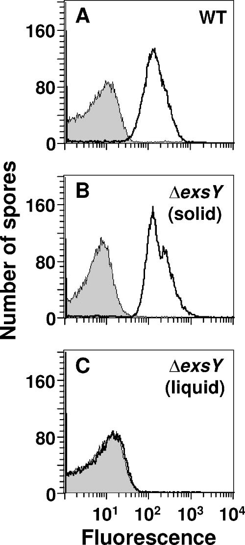
To determine if ExsY is required for the formation of the B. anthracis exosporium, we constructed a mutant version of the Sterne strain that is unable to produce this protein. The mutant, designated CLT325, contains a chromosomal mutation (ΔexsY) that precisely deletes the entire exsY gene, the only gene in its operon according to genome annotation (11), and replaces it with a spectinomycin resistance cassette. Strain CLT325, and the Sterne strain as a wild-type control, was grown on solid and in liquid media and allowed to sporulate. Mature spores were harvested, purified, and examined for the presence of the exosporium by flow cytometry after being incubated with fluorescently labeled anti-BclA MAb EF12. The results showed that the anti-BclA MAb bound similarly and extensively to Sterne spores produced with either solid or liquid medium (Fig. 1A and data not shown) and to ΔexsY spores grown on solid medium (Fig. 1B), indicating the presence of the exosporium. In contrast, the anti-BclA MAb did not bind to ΔexsY spores grown in liquid medium (Fig. 1C), indicating the absence of the exosporium.
FIG. 1.
Analysis by flow cytometry of binding of the anti-BclA MAb EF12 to wild-type and ΔexsY spores of B. anthracis. Spores of the wild-type Sterne strain (WT) produced in liquid medium (A) and spores of strain CLT325 (ΔexsY) produced on solid (B) or in liquid (C) medium were treated with fluorescently labeled EF12 (unfilled histograms with thick outlines) or an equivalently labeled isotype control MAb (gray histograms).
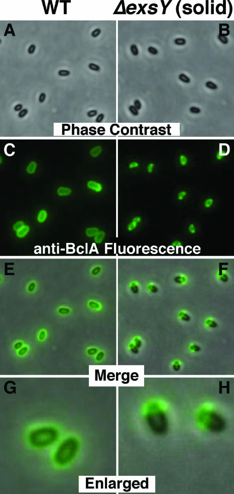
We further analyzed the Sterne and ΔexsY spores by phase-contrast and fluorescence microscopy (>100 spores inspected per sample), again following treatment with fluorescently labeled anti-BclA MAb EF12. All spores, whether grown on solid or in liquid medium, appeared similar by phase-contrast microscopy (Fig. 2A and B and data not shown). However, major differences were observed by fluorescence microscopy and from merged phase-contrast and fluorescence microscopic images. Sterne spores produced on solid or in liquid medium appeared uniformly and brightly fluorescent, as expected for spores surrounded by an exosporium (Fig. 2C, E, and G and data not shown). Surprisingly, fluorescent labeling of ΔexsY spores grown on solid medium was restricted to a polar cap-like region, which covered about one-third of the spore (Fig. 2D, F, and H). This result suggested that only a fragment of the exosporium was present on these spores. On the other hand, no labeling by the fluorescent anti-BclA MAb was observed with ΔexsY spores grown in liquid medium (data not shown). This result again indicates the lack of an exosporium on these spores.
FIG. 2.
Phase-contrast and fluorescence microscopic analysis of wild-type and ΔexsY spores of B. anthracis treated with fluorescently labeled anti-BclA MAb EF12. Spores of the wild-type Sterne strain (WT) were prepared in liquid medium (A, C, E, and G), and spores of strain CLT325 (ΔexsY) were produced on solid medium (B, D, F, and H). Phase-contrast images (A and B) and fluorescence images indicating the binding of EF12 to BclA (C and D) were used to produce merged (E and F) and enlarged merged (G and H) images.
Finally, we employed transmission electron microscopy to obtain high-resolution images of the Sterne and ΔexsY spores (>20 longitudinal spore sections inspected per sample). The Sterne spores produced on solid or in liquid medium possessed a fully developed exosporium, with a regular basal layer and hair-like nap (Fig. 3A and data not shown). As suggested by fluorescence microscopy, the transmission electron microscopic images of the ΔexsY spores grown on solid medium revealed a cap-like exosporium fragment covering one end of each spore (Fig. 3B). This fragment, which we call the “cap,” contained a normal hair-like nap but an irregular basal layer. As expected, the transmission electron microscopic images of the ΔexsY spores grown in liquid medium showed the complete loss of the exosporium from every spore examined (Fig. 3C). Other than the exosporium, the other layers of ΔexsY spores, whether exosporiumless or possessing a cap, appeared unaltered compared to wild-type Sterne spores.
FIG. 3.
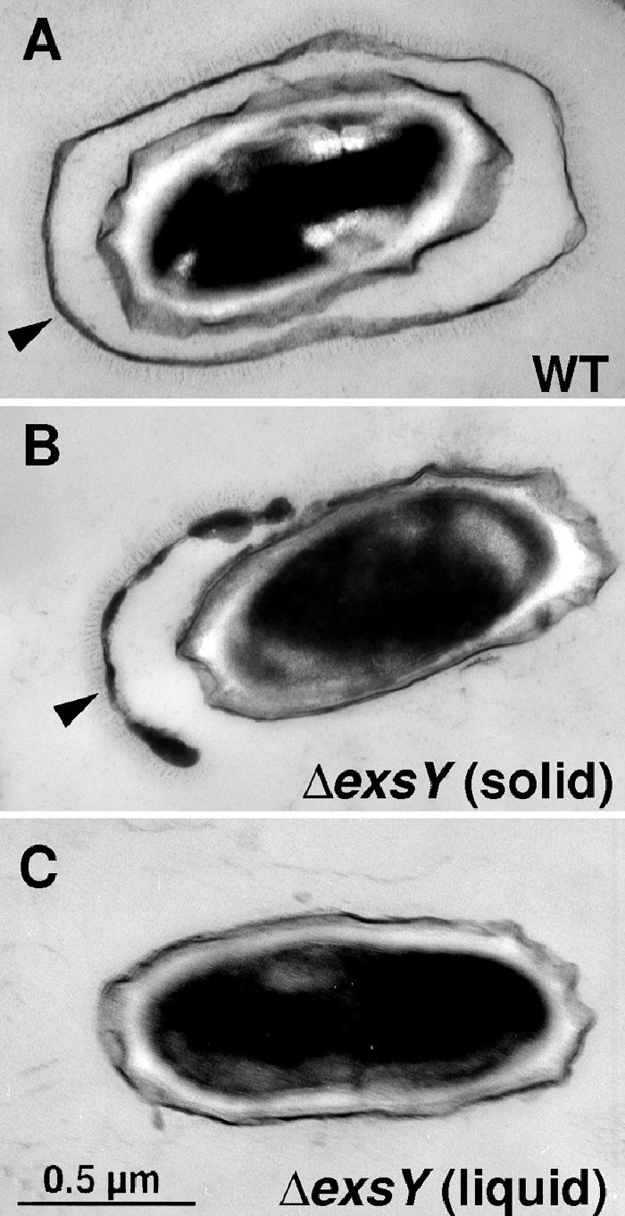
Transmission electron micrographs of wild-type and ΔexsY spores of B. anthracis. Thin sections of spores of the wild-type Sterne strain (WT) produced in liquid medium (A) and spores of strain CLT325 (ΔexsY) produced on solid (B) or in liquid (C) medium were examined. Arrowheads point to the exosporium (A) or cap-like exosporium fragment (B). The magnifications of all image are identical.
Exosporium development is arrested in sporulating ΔexsY cells.
To analyze the defect in exosporium development caused by the lack of ExsY, we examined sporulating cells of strain CLT325 (ΔexsY), and of the Sterne strain as a control, by transmission electron microscopy (>100 cells inspected per sample). Cells were grown and allowed to sporulate in liquid medium. Cells were harvested and fixed for examination hourly, starting 3 h and ending 9 h after the onset of sporulation (times hereafter are designated Tn, where n is the number of hours after the start of sporulation). Between T3 and T4, exosporium formation was first detected, and between T8 and T9, mature spores were released from the mother cell. At approximately T6, we first observed a clear difference in exosporium formation between the two strains. At this time, essentially all nascent spores within mother cells of the Sterne strain were completely enveloped by an exosporium, which contained a uniform basal layer and a normal hair-like nap (Fig. 4A). In contrast, the nascent spores within mother cells of the ΔexsY strain were only partially covered by a cap-like exosporium fragment (Fig. 4B). No further development of this fragment was detected at later time points, indicating a complete arrest of exosporium assembly. The T6 exosporium fragment, which contains a normal hair-like nap, appears to be the cap found on mature spores of the ΔexsY strain grown on solid medium (Fig. 3B). In every T6 cell of the ΔexsY strain, the exosporium fragment or cap covered the end of the spore near the middle of the mother cell. This result is consistent with previous studies showing that normal exosporium assembly begins at this location (20). The presence of a cap on all developing spores of the ΔexsY strain indicates that when spores of this strain are produced in liquid medium, they lose their caps. Prolonged shaking of the culture, or perhaps a presently unrecognized activity unique to liquid cultures, may dislodge the caps. Although spores of the ΔexsY strain do not assemble a complete exosporium, the other layers of these spores appear normal in sporangia (and also in mature spores, as indicated above).
FIG. 4.
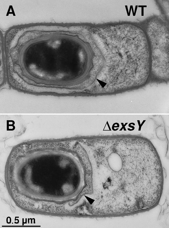
Transmission electron micrographs of sporulating cells of wild-type and ΔexsY strains of B. anthracis. Thin sections of sporulating (T6) cells of the wild-type (WT) Sterne strain (A) and strain CLT325 (ΔexsY) (B) were examined. Arrowheads point to the exosporium (A) or cap-like exosporium fragment (B). The magnifications of both images are identical.
An exosporium sublayer is revealed on ΔexsY spores.
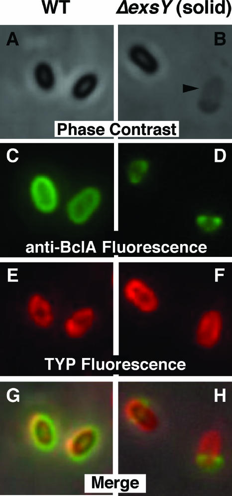
We previously identified a peptide, with the sequence TYPLPIR, that is capable of selectively binding to B. anthracis spores (37). Preliminary cross-linking experiments suggested that this peptide binds to ExsY (D. D. Williams and C. L. Turnbough, Jr., unpublished data). Therefore, we used fluorescence microscopy to examine the ability of a TYPLPIR peptide-PE conjugate (TYP-PE) to bind to purified ΔexsY spores. We observed no binding to spores prepared in liquid medium but did observe uniform and extensive binding to the surface of essentially every spore prepared on solid medium (data not shown). The latter result demonstrated that ExsY is not the target, or at least not the sole target, of TYP-PE binding, and it suggested that the site of TYP-PE binding on cap-containing ΔexsY spores is a fragile spore structure. To examine TYP-PE binding to spores with minimal damage to the presumed fragile structure, we prepared ΔexsY spores on solid medium without purification (i.e., spores were gently washed from plates with water). Sterne spores were prepared identically to serve as a wild-type control. A sample of each unpurified spore preparation was treated with both TYP-PE and Alexa Fluor 488-labeled anti-BclA MAb EF12 and then examined by phase-contrast and fluorescence microscopy (>100 spores inspected per sample).
Inspection by phase-contrast microscopy revealed refractile (i.e., phase bright) spores with a normal appearance in each spore prep (Fig. 5A and B and data not shown); however, in the preparation of ΔexsY spores, we also found an occasional spore-free sacculus (or sac) of approximately the size of the exosporium (Fig. 5B). Fluorescence staining by the anti-BclA MAb indicated a complete exosporium on Sterne spores (Fig. 5C and data not shown) and caps on all spores and nearly all sacs found in the ΔexsY spore preparation (Fig. 5D and data not shown). These assignments were confirmed by merging the phase-contrast images (Fig. 5A and B) with the corresponding anti-BclA fluorescence images (Fig. 5C and D) (data not shown). Fluorescence staining by TYP-PE indicated binding to the entire surfaces of Sterne spores, ΔexsY spores, and the sacs in the ΔexsY spore preparation (Fig. 5E and F and data not shown). Merged images of the fluorescence staining by TYP-PE and the anti-BclA MAb (which also include the phase-contrast images) indicated that the sacs represented a sublayer of the exosporium just under the outermost BclA-containing material in Sterne spores (Fig. 5G) and just under the caps of ΔexsY spores and sacs (Fig. 5H). Previous studies have suggested that the exosporium is composed of four closely packed lamellae (7), and the sublayer stained by TYP-PE may correspond to one of these.
FIG. 5.
Phase-contrast and fluorescence microscopic analysis of B. anthracis wild-type and ΔexsY spores treated with both Alexa Fluor 488-labeled anti-BclA MAb EF12 and TYP-PE. Spores of the wild-type Sterne (WT) and ΔexsY strains were produced on solid medium and harvested without purification. The phase-contrast images show two spores in the wild-type preparation (A) and one spore and one sac (indicated by an arrowhead) in the ΔexsY preparation (B). Fluorescence images show anti-BclA MAb staining (C and D) and TYP-PE staining (E and F) of the same material shown in the phase-contrast images. The corresponding phase-contrast, anti-BclA MAb fluorescence, and TYP-PE fluorescence images were merged to indicate overlapping staining (G and H).
When the ΔexsY spore preparation was stained with TYP-PE, many clusters of fragments of sac-like material were observed (data not shown), indicating that the sacs were easily broken and presumably corresponded to the fragile TYP-PE-binding material lost from ΔexsY spores grown in liquid medium. On the other hand, the same sacs within the exosporium of Sterne spores were extremely stable and apparently never sloughed from the spore. This observation is again consistent with the sac being an internal sublayer within the exosporium.
Exosporium proteins are lost from ΔexsY spores produced in liquid medium.
Sporulation of strain CLT325 (ΔexsY) in liquid medium produces mature spores that have shed all observable exosporium (including the cap and sac) but maintain the other layers of the spore in an apparently normal state. Consequently, these spores are likely to be devoid of proteins incorporated solely into the exosporium and deficient in proteins incorporated in part into this layer. Presently, about a dozen proteins are known or suspected to be incorporated into the exosporium, and we have rapid and reliable assays for about half of these. To further analyze the localization of the latter group of proteins, we compared their levels in purified wild-type Sterne and exosporiumless ΔexsY spores. Proteins were extracted from an equal number of Sterne and ΔexsY spores by boiling in sample buffer and then were separated by SDS-PAGE. The gels were either stained with Coomassie brilliant blue or used for immunoblotting.
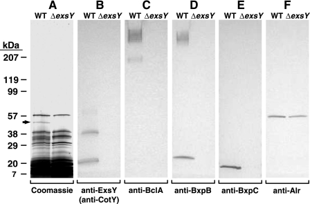
The Coomassie-stained gel was used to detect the putative exosporium protein inosine-uridine-preferring nucleoside hydrolase (IUNH). Monomeric IUNH, which has a mass of 36,276 Da, migrates with an apparent mass of approximately 50 kDa due to its low pI relative to those of protein standards. An essentially pure band of IUNH was readily detected in proteins extracted from Sterne spores (Fig. 6A). The identification and purity of the IUNH band were established by tryptic digestion and sequencing of the resulting fragments by tandem mass spectrometry (15; C. T. Steichen and C. L. Turnbough, Jr., unpublished data). In contrast, no IUNH was detectable in the proteins extracted from the ΔexsY spores (Fig. 6A). IUNH was not detected in this extract even when a severalfold larger sample was analyzed.
FIG. 6.
Detection of proteins extracted from Sterne (WT) and exosporiumless ΔexsY spores prepared in liquid medium. Proteins were separated by SDS-PAGE and visualized by staining with Coomassie brilliant blue (A) or detected by immunoblotting with mouse MAbs G9-3 (anti-ExsY/CotY) (B), EF12 (anti-BclA) (C), 10-23-4 (anti-BxpB) (D), FH6-1 (anti-BxpC) (E), and AR-1 (anti-Alr) (F). The arrow in panel A indicates the band corresponding to IUNH. The gel locations and masses (in kDa) of Bio-Rad protein standards are indicated on the left.
The relative levels of six other exosporium proteins were determined by immunoblotting using previously described mouse MAbs EF12 (anti-BclA), 10-23-4 (anti-BxpB), and FH6-1 (anti-BxpC) and mouse MAbs G9-3 (anti-ExsY) and AR-1 (anti-Alr), which were produced and characterized in this study, as described in Materials and Methods. In immunoblots, all MAbs except G9-3 react specifically with their designated target proteins, as either recombinant or spore proteins (data not shown). G9-3 reacts equally well with ExsY and its homologue CotY, again as either recombinant or spore proteins; for example, G9-3 reacts with CotY extracted from cap-containing ΔexsY spores and with ExsY extracted from ΔcotY spores (data not shown). ExsY and CotY have similar masses (i.e., 16,146 and 16,842 Da, respectively) and migrate as a doublet band during SDS-PAGE (data not shown). The immunoblot of Sterne spore proteins with G9-3 as the probe revealed doublet bands with masses expected for monomers, dimers, and trimers of ExsY and CotY (Fig. 6B). The same pattern of oligomerization was observed with rExsY and rCotY (data not shown). The apparent absence of ExsY and CotY in >250-kDa complexes is discussed below. The immunoblot of ΔexsY spore proteins with G9-3 failed to detect either ExsY or CotY (Fig. 6B). The immunoblots with EF12 (anti-BclA), 10-23-4 (anti-BxpB), and FH6-1 (anti-BxpC) as probes yielded similar results (Fig. 6C, D, and E). The blots showed the presence of BclA, BxpB, and BxpC in extracts of Sterne spores but failed to detect these proteins in extracts of ΔexsY spores. When extracted from Sterne spores, BclA and BxpB were included in large part in >250-kDa complexes (Fig. 6C and D), as previously reported (29), and a significant fraction of BxpB (17,331 Da) and all BxpC (14,379 Da) migrated as monomeric proteins (Fig. 6D and E). ExsY, CotY, BclA, BxpB, and BxpC were not detected in ΔexsY spore extracts, even when severalfold larger samples were analyzed. Thus, BclA, BxpB, BxpC, and CotY, along with IUNH, appear to be lost when the exosporium is shed from the ΔexsY spores.
In contrast, the immunoblot with AR-1 (which reacts with the spore-associated Alr protein but not with a second, vegetative-cell alanine racemase of B. anthracis) detected monomeric (43,662 Da) Alr in extracts of both Sterne and ΔexsY spores. However, the level of Alr from ΔexsY spores was reproducibly about half that from Sterne spores, as determined by densitometry (Fig. 6F and data not shown). Thus, Alr appears to be present in the exosporium plus at least one other extractable spore location, or alternatively, Alr is normally found solely in the exosporium but is aberrantly localized in ΔexsY spores.
It should be noted that the failure to detect BclA, BxpB, BxpC, and CotY in extracts of exosporiumless ΔexsY spores was not due to an inability to synthesize these proteins in sporulating ΔexsY cells. Using fluorescence microscopy and fluorescently labeled MAbs as probes, we detected high levels of BclA, BxpB, BxpC, and CotY in the caps of ΔexsY spores grown on solid medium (Fig. 2 and data not shown). Additionally, fluorescence microscopy with the fluorescently labeled MAb AR-1 detected Alr on the surfaces of Sterne spores, confirming the presence of this protein in the exosporium (data not shown).
The ΔexsY mutation does not affect cell growth, efficiency of spore production, or spore resistance.
To characterize the physiological effects of the ΔexsY mutation, we compared the growth rates, cell yields, and sporulation efficiencies of cultures of the Sterne and CLT325 (ΔexsY) strains grown in liquid DSM at 37°C with shaking. We observed no significant differences between the two cultures, with both growing with maximum doubling times of ∼25 min and sporulating with >95% efficiency (data not shown). Bacillus spores are resistant to heat, lysozyme, and organic solvents, such as chloroform, methanol, and phenol. Using standard protocols (19), we compared the levels of resistance to these treatments of purified preparations of Sterne and CLT325 (ΔexsY) spores produced in liquid DSM. We found no significant difference in the survival rates of Sterne and ΔexsY spores (data not shown).
The ΔexsY mutation does not affect the efficiency of spore germination but can alter the process of outgrowth.
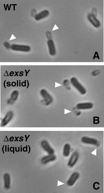
We also measured the effects of the ΔexsY mutation on spore germination and outgrowth. Spores of either the Sterne or CLT325 (ΔexsY) strain were dried on a coverslip, which was placed in a microscope chamber maintained at 37°C. After allowing the coverslip to warm to 37°C, the chamber was filled with prewarmed (37°C) growth medium containing RPMI 1640 with l-glutamine (Cellgro) and 0.2% brain heart infusion medium (Difco) (32). Spore germination and outgrowth were monitored by phase-contrast microscopy and time-lapse photography as previously described (29). The marker for germination was the change in spore appearance from refractile to phase dark, which occurs upon rehydration of the core and lysis of the cortex (21). The marker for outgrowth was the popping of the vegetative cell from its exosporium-containing “shell” (29). Previous studies indicated that this shell also includes the outer layer of the spore coat (16). Purified spores produced with both solid and liquid media were examined.
For all preparations, ≥95% of the spores germinated between 3 and 20 min of incubation in the growth medium. Most spores that did not germinate within this time period remained phase bright for the duration of the experiment (i.e., 90 min). The median time for germination of ΔexsY spores produced on solid or in liquid medium was approximately 6 min. The median time for germination of Sterne spores produced on solid or in liquid medium varied from 6 to 9 min. This variation was not related to the method of preparation but to differences observed with spores prepared under the same conditions. The source of this variation is unknown. For all spore preparations, essentially all outgrowth occurred between 35 and 60 min of incubation. Although it was not possible to reliably compare the median times of outgrowth for the four spore preparations due to a lack of synchrony, these preparations exhibited interesting differences during outgrowth. Both Sterne preparations outgrew identically, with popping of the vegetative cell from its shell within 10 s (i.e., the time between photographic images). Typically, this popping resulted in the complete escape of the outgrowing cell from its shell (Fig. 7A and data not shown). Although the shell often remained close to the outgrowing cell immediately after popping, a clear separation between them generally occurred within a few minutes. The ΔexsY spores produced on solid medium (i.e., cap-containing spores) exhibited popping essentially identical to that observed with Sterne spores (Fig. 7B). With these ΔexsY spores, the cap could be seen as a dark region on one end of the shell left behind by the outgrowing cell (Fig. 7B). This shell presumably contains the sac, perhaps other elements of the exosporium basal layer, and the outer spore coat. In contrast, the ΔexsY spores produced in liquid medium (i.e., devoid of all detectable exosporium) did not pop during outgrowth. Instead, the outgrowing cell gradually emerged from the shell, which presumably contained only the outer spore coat. Typically, this shell was only partially dislodged from the outgrowing cell (Fig. 7C). Apparently, complete escape of the outgrowing cell from its shell requires a more forceful exit.
FIG. 7.
Analysis of outgrowth of Sterne and ΔexsY spores. Wild-type Sterne (WT) spores produced in liquid medium (A) and ΔexsY spores produced on solid (B) or in liquid (C) medium were allowed to germinate and outgrow in growth medium for 55 min at 37°C and then examined by phase-contrast microscopy. White arrowheads indicate some of the shells that encapsulated the outgrowing cells.
DISCUSSION
The results of this study demonstrate that ExsY plays a critical role in the formation of the exosporium of B. anthracis. The absence of ExsY during sporulation causes an arrest in exosporium formation after the assembly of an exosporium fragment called the cap. The cap covers one end and approximately one-third of the forespore. The cap is assembled on the end of the forespore that is near the middle of the mother cell, which is also the site at which normal exosporium assembly is initiated (20). Thus, the cap presumably represents an early stage in exosporium development. The cap produced in the absence of ExsY contains a normal hair-like nap but an irregular basal layer, as judged from electron micrographs of purified spores. Therefore, ExsY is required not only to complete exosporium assembly after cap formation but also to form a normal cap. These requirements may reflect a checkpoint during exosporium development for proper cap formation or the absence of an essential structural element for continued exosporium assembly.
Although ExsY may be incorporated into a normal cap, its structural role in cap formation appears to be less important than its role in the assembly of the last two-thirds of the exosporium. Perhaps there is another protein that can substitute for ExsY early in exosporium assembly but not later in this process. A potential surrogate for ExsY during cap formation is CotY, which is 85% identical to ExsY and is present in cap-containing ΔexsY spores. The cotY gene (actually the cotY-bxpB operon) is apparently transcribed from a promoter recognized by the early mother cell sigma factor σE (29). The exsY gene (or exsY single-gene operon) appears to be transcribed from a promoter recognized by the late mother cell sigma factor σK (i.e., a σK-like promoter sequence is located 40 bp upstream of the exsY gene). Accordingly, CotY could be synthesized early in sporulation and incorporated into a nascent cap, while ExsY could be synthesized later and participate as an essential structural element for the last two-thirds of the exosporium. Confirmation of such a model will require the demonstration that normal exosporium assembly is discontinuous and/or that the exosporium contains segments with different protein compositions.
Previous studies demonstrated that ExsY is present in a high-molecular-mass complex that also contains BclA, BxpB, and possibly other proteins. This complex is highly stable and migrates with an apparent mass of >250 kDa during SDS-PAGE (24, 29). These observations suggested that ExsY plays an important role in exosporium assembly as part of the >250-kDa complex. However, the results of this study indicate that nearly all ExsY extracted from spores is in the form of monomers, dimers, and trimers. Apparently, only a small fraction of ExsY is stably incorporated into the >250-kDa complexes, which we confirmed by immunoblotting with the anti-ExsY/CotY MAb G9-3 and large amounts of extracted spore proteins (data not shown). Thus, the primary role of ExsY in exosporium assembly may not include the formation of highly stable complexes with BclA and BxpB.
Spores produced on solid medium by strain CLT325 (ΔexsY), i.e., in the absence of ExsY, retain the cap even after purification. Staining with a fluorescent conjugate of the spore binding peptide TYPLPIR (i.e., TYP-PE) and inspection by fluorescence microscopy revealed that these ΔexsY spores possess a layer called the sac. The sac appears to be a sublayer of the exosporium that underlies the cap while also covering the remainder of the spore. In wild-type spores, the sac appears to underlie the outermost BclA-containing sublayer of the exosporium. Thus, the sac may correspond to an inner layer of the four closely packed lamellae that were previously reported to make up the basal layer of the exosporium (7). The sac appears to be stable in wild-type and cap-containing ΔexsY spores. However, when the sac is shed from ΔexsY spores, it is extremely fragile. Spores produced in liquid medium by strain CLT325 lose the cap and also the sac. Whether these losses reflect physical damage caused by shaking of the sporulating culture or an activity uniquely associated with the liquid culture remains to be determined. Although the sac layer was readily observed on cap-containing ΔexsY spores by fluorescence microscopy, electron micrographs of these spores or of sporangia in which these spores were being produced did not reveal an exosporium-like layer (i.e., the sac) surrounding the entire spore. Possibly, the sac is refractile to the staining employed for electron microscopy or the sac was destroyed during sample preparation. The protein composition of the sac is presently under investigation.
The observation that purified CLT325 (ΔexsY) spores produced on solid medium retain their caps suggests a connection between the exosporium and the rest of the spore. Such connections could direct the assembly of the exosporium around the developing forespore. In sporulating ΔexsY cells, both ends of the cap bend towards, and are close to, the rest of the forespore (Fig. 4B). Perhaps these clamp-like ends represent the proposed connections. These or related connections could persist in the mature spore and provide structural stability. At present, no connections between the exosporium and other layers of the mature spore have been detected.
The ΔexsY spores produced in liquid medium lacked an exosporium and presumably had lost the proteins used to assemble this layer. Therefore, we examined these spores for the loss of several confirmed and putative exosporium proteins for which we have good assays. Our results showed that BclA, BxpB, BxpC, CotY, and IUNH were undetectable, indicating that these proteins are present solely in the exosporium. The level of another putative exosporium protein, the spore-associated protein Alr, was reduced to approximately 50% of that found on wild-type spores. This result suggests that Alr is present in the exosporium and at least one other spore location or that Alr is aberrantly localized in ΔexsY spores. We are currently comparing the protein contents of wild-type and ΔexsY spores by using proteomic and other analytical techniques to further characterize exosporium proteins.
Deletion of the exsY gene did not significantly alter cell growth, the efficiency of sporulation and germination, or the resistance of spores to heat, lysozyme, and organic solvents. These observations indicate a lack of involvement in these activities of exosporium features lost from ΔexsY spores. The median times for germination and outgrowth of ΔexsY spores, whether produced on solid or in liquid medium, were either the same or a few minutes shorter than those observed for similarly prepared wild-type spores. This result was unexpected because ΔexsY spores, at least those grown in liquid medium and devoid of an exosporium, lack BxpB. This protein was recently shown to delay spore germination and outgrowth (29). This apparent discrepancy will require further investigation. Our observations of outgrowth of ΔexsY spores provided a surprise. We expected that germinating ΔexsY spores would not exhibit the popping phenomenon observed when germinating wild-type spores suddenly escape their exosporium encasement. However, ΔexsY spores containing a cap exhibited popping essentially identical to that of wild-type spores. Apparently, the cap-containing ΔexsY spores are surrounded by a structure, presumably one that includes the sac, that also abruptly ruptures following sufficient enlargement of the germinating spore. In contrast, exosporiumless ΔexsY spores did not exhibit popping during outgrowth but exhibited a gradual emergence of the outgrowing cell from a shell that presumably contained only spore coat material. It appears that this material does not constitute a persistent physical barrier to spore germination and outgrowth.
The exosporium is conserved among all pathogenic Bacillus species, including B. anthracis, the opportunistic human pathogen B. cereus, and the insect pathogen B. thuringiensis. The high energetic cost of maintaining an elaborate exosporium is presumably offset by survival advantages that contribute to spore viability and virulence. However, these advantages remain to be established. Recent studies have shown that the mechanical removal of the exosporium from wild-type B. anthracis spores results in increased killing by murine macrophages (12) and by nitric oxide (23). It will be interesting to determine whether exosporiumless ΔexsY spores exhibit similar susceptibilities. In addition, these exosporiumless spores should be useful tools for new studies to evaluate the roles of the exosporium, perhaps in spore survival in harsh environments or during interactions with the cells of the mammalian immune system.
Acknowledgments
We thank Leigh Millican of the UAB High Resolution Imaging Facility for invaluable assistance with electron microscopy. Mass spectrometry was performed by Marion Kirk and Landon Wilson in the UAB Comprehensive Cancer Center Mass Spectrometry Shared Facility. We thank Chris Steichen for his help and advice.
This work was supported by NIH grants AI057699 and AI50566.
Footnotes
Published ahead of print on 25 August 2006.
REFERENCES
- 1.Boydston, J. A., P. Chen, C. T. Steichen, and C. L. Turnbough, Jr. 2005. Orientation within the exosporium and structural stability of the collagen-like glycoprotein BclA of Bacillus anthracis. J. Bacteriol. 187:5310-5317. [DOI] [PMC free article] [PubMed] [Google Scholar]
- 2.Brittingham, K. C., G. Ruthel, R. G. Panchal, C. L. Fuller, W. J. Ribot, T. A. Hoover, H. A. Young, A. O. Anderson, and S. Bavari. 2005. Dendritic cells endocytose Bacillus anthracis spores: implications for anthrax pathogenesis. J. Immunol. 174:5545-5552. [DOI] [PubMed] [Google Scholar]
- 3.Daubenspeck, J. M., H. Zeng, P. Chen, S. Dong, C. T. Steichen, N. R. Krishna, D. G. Pritchard, and C. L. Turnbough, Jr. 2004. Novel oligosaccharide side-chains of the collagen-like region of BclA, the major glycoprotein of the Bacillus anthracis exosporium. J. Biol. Chem. 279:30945-30953. [DOI] [PubMed] [Google Scholar]
- 4.Foster, S. J., and D. L. Popham. 2002. Structure and synthesis of cell wall, spore cortex, teichoic acids, S-layers, and capsules, p. 21-41. In A. L. Sonenshein, J. A. Hoch, and R. Losick (ed.), Bacillus subtilis and its closest relatives. From genes to cells. ASM Press, Washington, D.C.
- 5.Gerhardt, P. 1967. Cytology of Bacillus anthracis. Fed. Proc. 26:1504-1517. [PubMed] [Google Scholar]
- 6.Gerhardt, P., and S. H. Black. 1961. Permeability of bacterial spores. II. Molecular variables affecting solute permeation. J. Bacteriol. 82:750-760. [DOI] [PMC free article] [PubMed] [Google Scholar]
- 7.Gerhardt, P., and E. Ribi. 1964. Ultrastructure of the exosporium enveloping spores of Bacillus cereus. J. Bacteriol. 88:1774-1789. [DOI] [PMC free article] [PubMed] [Google Scholar]
- 8.Green, B. D., L. Battisti, T. M. Koehler, C. B. Thorne, and B. E. Ivins. 1985. Demonstration of a capsule plasmid in Bacillus anthracis. Infect. Immun. 49:291-297. [DOI] [PMC free article] [PubMed] [Google Scholar]
- 9.Guidi-Rontani, C., M. Levy, H. Ohayon, and M. Mock. 2001. Fate of germinated Bacillus anthracis spores in primary murine macrophages. Mol. Microbiol. 42:931-938. [DOI] [PubMed] [Google Scholar]
- 10.Henriques, A. O., and C. P. Moran, Jr. 2000. Structure and assembly of the bacterial endospore coat. Methods 20:95-110. [DOI] [PubMed] [Google Scholar]
- 11.Kanehisa, M., S. Goto, M. Hattori, K. F. Aoki-Kinoshita, M. Itoh, S. Kawashima, T. Katayama, M. Araki, and M. Hirakawa. 2006. From genomics to chemical genomics: new developments in KEGG. Nucleic Acids Res. 34:D354-D357. [DOI] [PMC free article] [PubMed] [Google Scholar]
- 12.Kang, T. J., M. J. Fenton, M. A. Weiner, S. Hibbs, S. Basu, L. Baillie, and A. S. Cross. 2005. Murine macrophages kill the vegetative form of Bacillus anthracis. Infect. Immun. 73:7495-7501. [DOI] [PMC free article] [PubMed] [Google Scholar]
- 13.Kearney, J. F., R. Barletta, Z. S. Quan, and J. Quintans. 1981. Monoclonal vs. heterogeneous anti-H-8 antibodies in the analysis of the anti-phosphorylcholine response in BALB/c mice. Eur. J. Immunol. 11:877-883. [DOI] [PubMed] [Google Scholar]
- 14.Lai, E. M., N. D. Phadke, M. T. Kachman, R. Giorno, S. Vazquez, J. A. Vazquez, J. R. Maddock, and A. Driks. 2003. Proteomic analysis of the spore coats of Bacillus subtilis and Bacillus anthracis. J. Bacteriol. 185:1443-1454. [DOI] [PMC free article] [PubMed] [Google Scholar]
- 15.Mann, M., R. C. Hendrickson, and A. Pandey. 2001. Analysis of proteins and proteomes by mass spectrometry. Annu. Rev. Biochem. 70:437-473. [DOI] [PubMed] [Google Scholar]
- 16.Moberly, B. J., F. Shafa, and P. Gerhardt. 1966. Structural details of anthrax spores during stages of transformation into vegetative cells. J. Bacteriol. 92:220-228. [DOI] [PMC free article] [PubMed] [Google Scholar]
- 17.Mock, M., and A. Fouet. 2001. Anthrax. Annu. Rev. Microbiol. 55:647-671. [DOI] [PubMed] [Google Scholar]
- 18.Nicholson, W. L., N. Munakata, G. Horneck, H. J. Melosh, and P. Setlow. 2000. Resistance of Bacillus endospores to extreme terrestrial and extraterrestrial environments. Microbiol. Mol. Biol. Rev. 64:548-572. [DOI] [PMC free article] [PubMed] [Google Scholar]
- 19.Nicholson, W. L., and P. Setlow. 1990. Sporulation, germination and outgrowth, p. 391-450. In C. R. Harwood and S. M. Cutting (ed.), Molecular biological methods for Bacillus. John Wiley & Sons, Ltd., West Sussex, England.
- 20.Ohye, D. F., and W. G. Murrell. 1973. Exosporium and spore coat formation in Bacillus cereus T. J. Bacteriol. 115:1179-1190. [DOI] [PMC free article] [PubMed] [Google Scholar]
- 21.Popham, D. L., J. Helin, C. E. Costello, and P. Setlow. 1996. Muramic lactam in peptidoglycan of Bacillus subtilis spores is required for spore outgrowth but not for spore dehydration or heat resistance. Proc. Natl. Acad. Sci. USA 93:15405-15410. [DOI] [PMC free article] [PubMed] [Google Scholar]
- 22.Priest, F. G. 1993. Systematics and ecology of Bacillus, p. 3-16. In A. L. Sonenshein, J. A. Hoch, and R. Losick (ed.), Bacillus subtilis and other gram-positive bacteria. Biochemistry, physiology, and molecular biology. American Society for Microbiology, Washington, D.C.
- 23.Raines, K. W., T. J. Kang, S. Hibbs, G. L. Cao, J. Weaver, P. Tsai, L. Baillie, A. S. Cross, and G. M. Rosen. 2006. Importance of nitric oxide synthase in the control of infection by Bacillus anthracis. Infect. Immun. 74:2268-2276. [DOI] [PMC free article] [PubMed] [Google Scholar]
- 24.Redmond, C., L. W. Baillie, S. Hibbs, A. J. Moir, and A. Moir. 2004. Identification of proteins in the exosporium of Bacillus anthracis. Microbiology 150:355-363. [DOI] [PubMed] [Google Scholar]
- 25.Rider, T. H., M. S. Petrovick, F. E. Nargi, J. D. Harper, E. D. Schwoebel, R. H. Mathews, D. J. Blanchard, L. T. Bortolin, A. M. Young, J. Chen, and M. A. Hollis. 2003. A B cell-based sensor for rapid identification of pathogens. Science 301:213-215. [DOI] [PubMed] [Google Scholar]
- 26.Sambrook, J., E. F. Fritsch, and T. Maniatis. 1989. Molecular cloning: a laboratory manual, 2nd ed. Cold Spring Harbor Laboratory, Cold Spring Harbor, N.Y.
- 27.Setlow, P. 2003. Spore germination. Curr. Opin. Microbiol. 6:550-556. [DOI] [PubMed] [Google Scholar]
- 28.Steichen, C., P. Chen, J. F. Kearney, and C. L. Turnbough, Jr. 2003. Identification of the immunodominant protein and other proteins of the Bacillus anthracis exosporium. J. Bacteriol. 185:1903-1910. [DOI] [PMC free article] [PubMed] [Google Scholar]
- 29.Steichen, C. T., J. F. Kearney, and C. L. Turnbough, Jr. 2005. Characterization of the exosporium basal layer protein BxpB of Bacillus anthracis. J. Bacteriol. 187:5868-5876. [DOI] [PMC free article] [PubMed] [Google Scholar]
- 30.Stopa, P. J. 2000. The flow cytometry of Bacillus anthracis spores revisited. Cytometry 41:237-244. [DOI] [PubMed] [Google Scholar]
- 31.Stragier, P., and R. Losick. 1996. Molecular genetics of sporulation in Bacillus subtilis. Annu. Rev. Genet. 30:297-341. [DOI] [PubMed] [Google Scholar]
- 32.Swiecki, M. K., M. W. Lisanby, C. L. Turnbough, Jr., and J. F. Kearney. 2006. Monoclonal antibodies for Bacillus anthracis spore detection and functional analyses of spore germination and outgrowth. J. Immunol. 176:6076-6084. [DOI] [PubMed] [Google Scholar]
- 33.Sylvestre, P., E. Couture-Tosi, and M. Mock. 2002. A collagen-like surface glycoprotein is a structural component of the Bacillus anthracis exosporium. Mol. Microbiol. 45:169-178. [DOI] [PubMed] [Google Scholar]
- 34.Sylvestre, P., E. Couture-Tosi, and M. Mock. 2005. Contribution of ExsFA and ExsFB proteins to the localization of BclA on the spore surface and to the stability of the Bacillus anthracis exosporium. J. Bacteriol. 187:5122-5128. [DOI] [PMC free article] [PubMed] [Google Scholar]
- 35.Todd, S. J., A. J. G. Moir, M. J. Johnson, and A. Moir. 2003. Genes of Bacillus cereus and Bacillus anthracis encoding proteins of the exosporium. J. Bacteriol. 185:3373-3378. [DOI] [PMC free article] [PubMed] [Google Scholar]
- 36.Weiner, M. A., and P. C. Hanna. 2003. Macrophage-mediated germination of Bacillus anthracis endospores requires the gerH operon. Infect. Immun. 71:3954-3959. [DOI] [PMC free article] [PubMed] [Google Scholar]
- 37.Williams, D. D., O. Benedek, and C. L. Turnbough, Jr. 2003. Species-specific peptide ligands for the detection of Bacillus anthracis spores. Appl. Environ. Microbiol. 69:6288-6293. [DOI] [PMC free article] [PubMed] [Google Scholar]
- 38.Williams, D. D., and C. L. Turnbough, Jr. 2004. Surface layer protein EA1 is not a component of Bacillus anthracis spores but is a persistent contaminant in spore preparations. J. Bacteriol. 186:566-569. [DOI] [PMC free article] [PubMed] [Google Scholar]


