
FIG. 4.
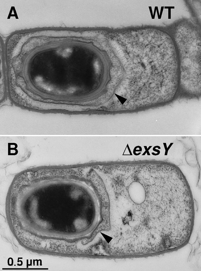
Transmission electron micrographs of sporulating cells of wild-type and ΔexsY strains of B. anthracis. Thin sections of sporulating (T6) cells of the wild-type (WT) Sterne strain (A) and strain CLT325 (ΔexsY) (B) were examined. Arrowheads point to the exosporium (A) or cap-like exosporium fragment (B). The magnifications of both images are identical.