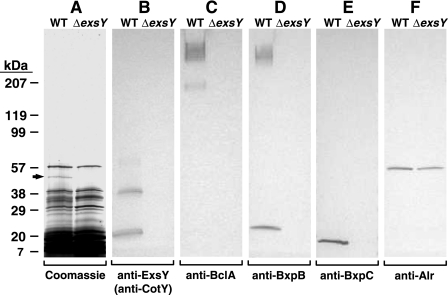
FIG. 6.
Detection of proteins extracted from Sterne (WT) and exosporiumless ΔexsY spores prepared in liquid medium. Proteins were separated by SDS-PAGE and visualized by staining with Coomassie brilliant blue (A) or detected by immunoblotting with mouse MAbs G9-3 (anti-ExsY/CotY) (B), EF12 (anti-BclA) (C), 10-23-4 (anti-BxpB) (D), FH6-1 (anti-BxpC) (E), and AR-1 (anti-Alr) (F). The arrow in panel A indicates the band corresponding to IUNH. The gel locations and masses (in kDa) of Bio-Rad protein standards are indicated on the left.

