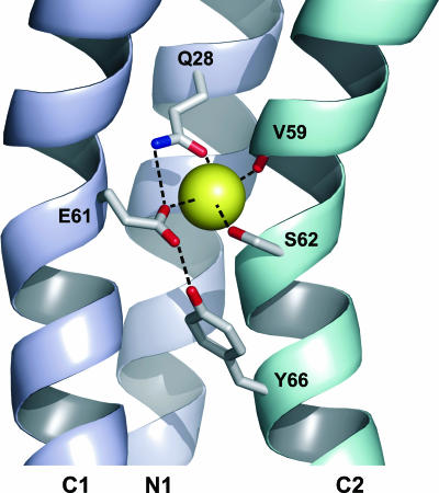
FIG. 5.
Structure of the C. paradoxum c11 ring Na+-binding site obtained from homology modeling in ribbon representation. Two c subunits are shown in different colors. Each subunit is formed by an inner (N1) and an outer (C1 and C2) helix. The view is normal to the external surface, with the ring axis being vertical. The amino acid side chains mentioned in the text are indicated in stick representation. The Na+ coordination and selected hydrogen bonds are indicated with dashed lines. The image was created with PyMOL (5).

