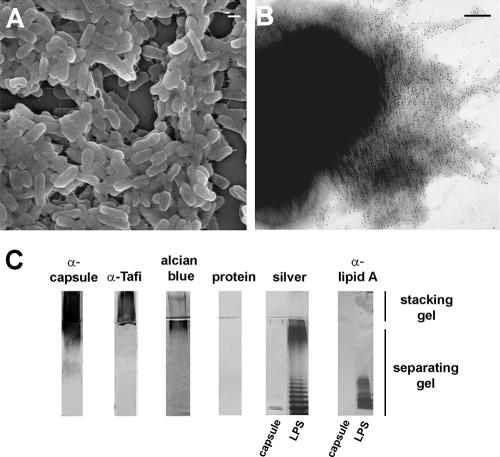
FIG. 1.
Salmonella EPS associated with the extracellular matrix. (A) Scanning electron micrograph of SE 3b cells embedded in the extracellular matrix (bar = 1 μm). (B) Transmission electron micrograph of SE 3b expressing the extracellular matrix immunostained with serum generated against the purified EPS (bar = 100 nm). (C) SDS-PAGE and immunoblot analysis of 10 μg of purified EPS from SE 3b ΔbcsA or 10 μg of LPS from Salmonella serovar Enteritidis (Sigma).

