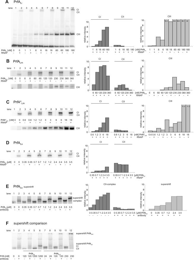
FIG. 3.
Determination of the binding affinity of the PrfA proteins with and without RNA polymerase to the PrfA-dependent promoter PhlyLm by EMSA. EMSAs were performed with purified PrfA proteins of L. ivanovii (PrfALi), L. monocytogenes (PrfALm and PrfA*Lm), and L. seeligeri (PrfALs) at various concentrations, 5 nM hly DNA promoter fragment of L. monocytogenes (PhlyLm), and 1.5 nM RNAP. CI (complex of DNA, RNAP, and PrfA), CII (complex of DNA and RNAP), and CIII (complex of DNA and PrfA) were quantified by using the ImageMaster program (Total Lab software, version 1.11; Amersham) and are shown in the graphs to the right. Lane 2 in each panel shows a PhlyLm control with L. monocytogenes RNAP. The intensity of this band is taken as 1, and all other values are normalized to it (values shown on y axis). The data shown here represent the results of one of three independently performed experiments. Supershift assays were performed with purified anti-PrfA antibodies (in a final concentration of 1:15) and increasing concentrations of PrfA protein (0.35, 0.7, 1.2, 2.4, and 3.5 μM PrfALs) (E), and a supershift comparison of PrfALs (120 and 1,200 nM) with PrfALm (24, 120, and 230 nM) was performed (F).

