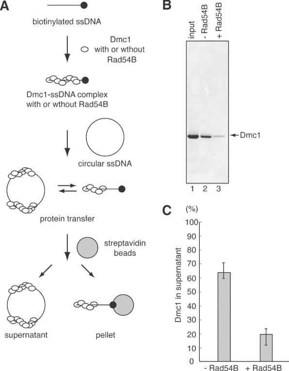
Figure 3.
Pull down assay for the protein transfer between ssDNA molecules. (A) A schematic diagram of the pull down assay. Dmc1 and biotinylated DNA form a complex in the absence or presence of Rad54B. Then, circular ssDNA is added as a competitor, and protein transfer occurs. Biotinylated DNA is immobilized on streptavidin beads, and the reaction mixture is divided into the beads and supernatant. (B) Dmc1 (5 µM) was incubated with SAT-120 (20 µM) labeled with biotin at the 5′ end in the absence (lane 2) or presence (lane 3) of Rad54B (200 nM), followed by an incubation with φX174 circular ssDNA (2 mM). After immobilization on streptavidin beads for 1 h at 4°C, the reaction mixture was divided into the beads and the supernatant by centrifugation. Then, one-tenth of the supernatant was fractionated on a 4–20% gradient SDS–PAGE gel. Lane 1 is one-tenth of the input protein. (C) Graphic representation of the experiments shown in (B). The amounts of Dmc1 within the supernatant are presented.

