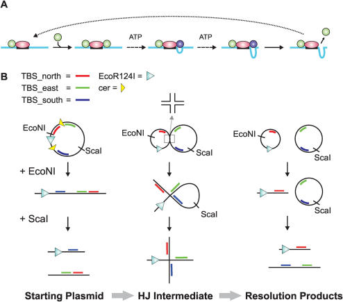
Figure 1.
DNA substrates for the analysis of junction bypass by a Type I restriction enzyme. (A) DNA translocation by a Type I restriction enzyme. DNA shown as a blue line with a restriction site as a black box; EcoR124I MTase shown as a pink oval; HsdR shown as a green (static) or blue (translocating) circle. For the sake of clarity, translocation on only one side of the site is shown. See main text for more details. (B) Generation of DNA junction substrates. E.coli RM40 cells were transformed with pLKS7 and recombination induced (see Materials and Methods for further details). Cells were harvested and the DNA purified as a mixture of starting plasmid (unrecombined pLKS7), HJ intermediate and resolution products (recombined circular products). Treatment with either EcoNI or EcoNI plus ScaI produced the substrates shown. The DNA was further purified using standard techniques, without separation of the different species. Approximate locations of the TFOs are given by the blue, red and green lines, respectively. More detailed information on spacings is given in Figures 3 and 5.

