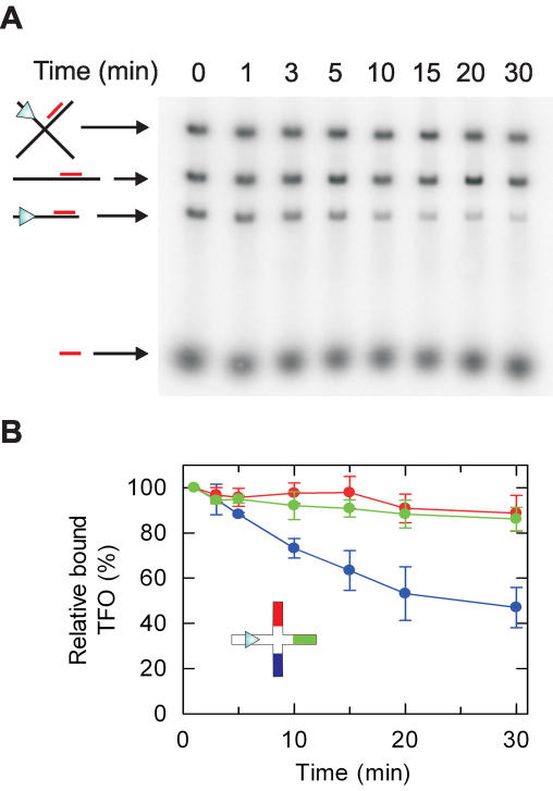
Figure 4.
Translocation on χ-substrate DNA at low HsdR concentrations. (A) Representative agarose gel showing separation of the different DNA produced by EcoNI/ScaI digestion and species bound by TFO_north. DNA (5 nM) was pre-bound with 32P-labelled TFO_north as described (11), and then treated with 1 nM HsdR and 40 nM MTase in buffer R for the times shown. Images were captured using a Molecular Dynamics Typhoon 9200 PhosphorImager and quantified using ImageQuant software. (B) TFO-binding to the χ-species was calculated relative to the 1 min sample (with the bound TFO at t = 60 s set to 100%). Each triplex was analysed in a separate reaction. Data represents the average of ≥2 independent experiments.

