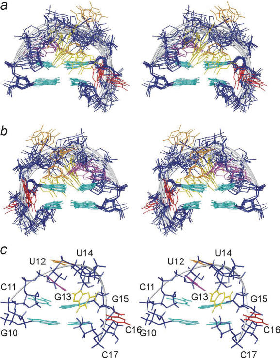
Figure 2.
Stereo views of an overlay of the 12 structures of the pseudo-triloop selected in step A2. (a) Viewed into the minor groove and (b) into the major groove. The sugar–phosphate backbone is coloured dark blue and the fold of the backbone is indicated as light grey tubes; colouring scheme of nucleobases is G10, C11, G15 and C17, light blue; U12, magenta; G13, yellow; U14, orange and C16, red. (c) The best structure as defined by the selection criteria.

