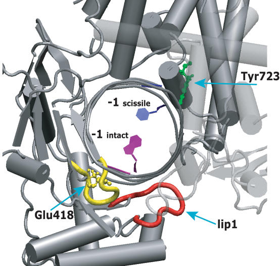
Figure 8.
Human topoisomerase I structure in complex with duplex DNA. The main chain of the 417–423 loop, contacting the intact strand of the lip 1 (represented in red), is shown in yellow. The mutated Glu418 and the catalytic Tyr723 residues are represented in ball and stick, in green and yellow colours, respectively. The −1 base of the scissile strand, strictly required for CPT binding, is coloured in blue; the −1 base of the intact strand, that contacts the 417–423 loop, is represented in purple. All other enzyme residues and DNA bases are represented in grey. Core subdomain III is partial transparent.

