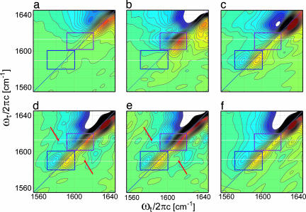
Fig. 4.
The 2D correlation spectra of GpA TM homodimers G79 (a), G*79 (b), and G**79 (c); heterodimers G*79 + G**79 (d) and (G*79 + G**79) − 0.25G**79 (e); and control sample G*94 + G**79 (f) in 5% SDS. The 13C 16O and 13C
 18O isotopomer diagonal regions are highlighted by rectangular boxes, and the arrows point to the cross-peaks.

