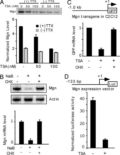
Fig. 2.
HDAC inhibitors require new protein synthesis to inhibit Mgn gene expression. Differentiated myotubes were used to investigate the effect of HDAC inhibition on Mgn gene expression. (A) Western blot analysis of Mgn protein levels in active (−TTX) and inactive (+TTX) myotubes treated with either buffer or TSA for 6 h. Graph below the blot shows quantification of Mgn protein levels shown on Western blot after normalization to total protein applied to the gel (n = 3). (B) New protein synthesis is required for Mgn RNA suppression by HDAC inhibitors. C2C12 myotubes were treated with buffer or NaB in the presence or absence of cycloheximide (CHX) for 6 h before isolating RNA for radioactive RT PCR and analysis on polyacrylamide gels (autoradiogram shown, Upper). (B Lower) Graph shows quantification of data shown in autoradiogram (n = 3). (C and D) Mgn promoter activity is suppressed by HDAC inhibitors. (C) C2C12 myotubes harboring an integrated 1-kb Mgn promoter driving GFP gene expression were treated with and without TSA and CHX for 12 h before quantifying GFP and actin RNA levels by real-time PCR (n = 3). GFP RNA level is normalized to actin RNA. (D) C2C12 cells were transfected with a 133-bp Mgn promoter-luciferase expression vector along with CMV-CAT for normalization. Transfected myotubes were treated with and without TSA for 12 h before cell harvest for luciferase and CAT assays (n = 3). Error bars are standard deviation.

