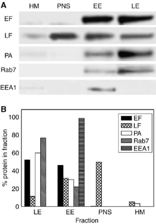
Figure 3.

Different subcellular localizations of anthrax LF and EF. HeLa cells were treated either with LF+PA (10 and 20 nM, respectively) or with EF+PA (10 and 20 nM, respectively) for 1 h at 37°C and were then lysed and separated into a supernatant (PNS), which includes the cell cytosol, and a membrane fraction that was fractionated on a sucrose density gradient. Three fractions were obtained: HM, heavy membranes, EE, early endosomes and LE, late endosomes. Ten microgram proteins of these fractions were subjected to SDS–PAGE, immunoblotted and developed as described in the Materials and methods. (A) Toxin and markers distribution among the different fractions in a representative experiment, while (B) show the corresponding quantification of the bands, which includes a normalization to the total amount of proteins of each of the four fractions examined. Rab7 is a marker of late endosomes and EEA1 is a marker of early endosomes. Note the largely different distribution of LF and EF, with LF being present mainly in the cell cytosol fraction and EF on endosomes. The significant amount of LF and EF still present in the early endosomal fraction is due to the fact that the experiment was performed without low-temperature synchronization of the binding step, under the same conditions required for imaging (see text).
