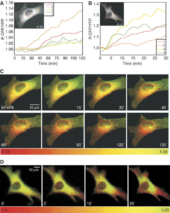
Figure 5.

Anthrax edema toxin creates c-AMP microdomains in HeLa cells. (A) HeLa cells expressing the cytosolic PKA-based probe cAMP fluorescence biosensor were treated with EF 10+PA 20 nM (time zero) and maintained in 2 ml of balanced salt solution at 37°C during microscopic observations. CFP/YFP ratios were measured in the indicated areas, identified with different colour contours: perinuclear regions (1, red trace; 2, orange trace) and cell periphery (3, yellow trace; 4, green trace). Notice the lower cAMP rising in the peripheral areas. (B) HeLa cell expressing the cAMP cytosolic probe treated with the B. pertussis CyaA adenylate cyclase toxin, which enters from the plasma membrane. Notice the faster rise of the ratiometic signal in the sub-plasma membrane areas identified by different colours, which are the same of those of the corresponding traces. (C, D) Pseudo-colour images, generated by CFP/YFP ratio imaging, of the intracellular cAMP at the given time points of the cell of (A) treated with PA+EF and of the cell of (B) treated with CyaA.
