
Figure 5.
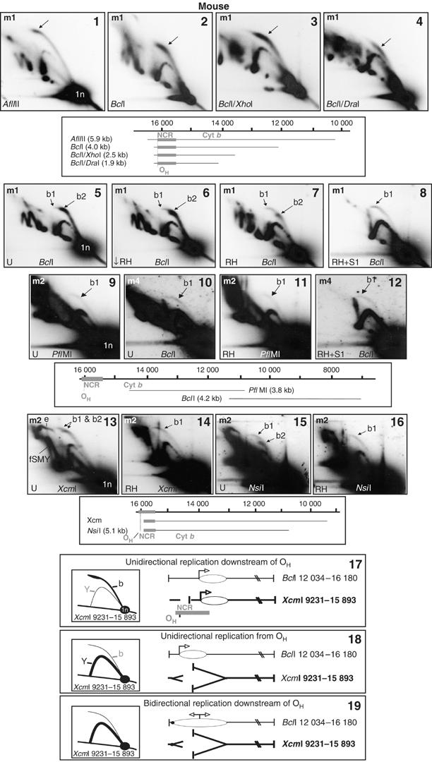
RNase H-sensitive ‘clubheaded' initiation arcs localize the origin of ribonucleotide-incorporating replication to the NCR. 2D-AGE analysis of NCR-containing fragments of mouse liver mtDNA, digested with the restriction enzymes and hybridized with probes as indicated, m1 (panels 1–8), m2 (panels 9, 11 and 13–16) or m4 (panels 10 and 12). Diagrams indicating the fragments detected are shown beneath the relevant gel panels. Panels 5–16 were either treated with low levels of RNase H (↓ RH, 0.1 U, 30 min, 37°C), or our standard treatment (RH, 1 U, 30 min, 37°C) or S1 nuclease (S1, 1 U, 60 s, 37°C) or else left untreated with nuclease (U). Mock RNase H treatments or very low doses of RNase H were indistinguishable from untreated samples (data not shown). Bubble arcs are arrowed: b1—RNase H-insensitive bubble arcs detected across the origin zone (Ori-Z); b2—RNase H-sensitive, clubheaded bubble arcs detected only in NCR-containing fragments. See also Supplementary Figure 5. Panels 13–16: a weak RNase H-sensitive initiation arc is associated with fragments of mouse liver mtDNA containing some of the NCR yet lacking OH. Probe m2 was applied to a 2D blot of XcmI digested mouse mtDNA treated without (panel 13) and with (panel 14) RNase H, revealing a 6.65 kb fragment (nt 9231–15 893) including >400 nts of the NCR. Similarly, NsiI fragment nt 10 765–15 909 yielded two initiation arcs (panel 15), only one of which was RNase H resistant (panel 16). The RNase H-sensitive, clubheaded bubble arcs of non OH-containing fragments, (panels 13 and 15), are compatible only with unidirectional replication initiating in the NCR downstream of OH (panel 17). Three panels (17–19) depict alternative hypothetical modes of replication initiation. Panel 17 represents unidirectional replication from the OH-distal end of the NCR; panel 18 shows unidirectional replication from OH; panel 19 represents bidirectional replication from the OH-distal end of the NCR. Of these three modes of initiation, only the one shown in panel 17 is consistent with the initiation arcs seen in panels 13 and 15, as indicated by the hypothetical 2D-AGE gel images shown alongside each model; b—bubble arc; Y—simple replication fork arc. This model, of a fraction of molecules initiating unidirectional replication at the cytochrome b gene end of the NCR, is also consistent with the detection of a cluster of H-strand 5′ ends mapping to the OH-distal end of the NCR (Figure 6(1)).