
Figure 2.
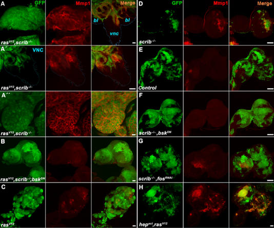
JNK-dependent expression of Mmp1 in rasV12, scrib−/− tumors. Third instar eye/antennal imaginal discs carrying GFP-marked clones (green) of the indicated genotypes were immunostained with an anti-Mmp1 antibody (red) and analyzed by confocal microscopy. (A) Strong upregulation of Mmp1 protein expression in rasV12, scrib−/− clones is seen not only in overgrown eye/antennal discs but also in cells invading distal parts of the brain such as the VNC (A, right). Magnification of the VNC with invading Mmp1-positive cells (A′). Mmp1 protein is enriched on surface of malignant rasV12, scrib−/− cells (A″). (B) Mmp1 expression is lost when JNK activity is suppressed by the expression of BskDN in rasV12, scrib−/− clones. Note that the clonal tissue still overgrows the entire eye/antennal imaginal disc. (C–E) While significantly smaller, scrib−/− clones show prominent Mmp1 staining (D) compared to the bigger wild-type control (E) or rasV12 (C) clones. (F, G) Mmp1 expression in scrib−/− clones is lost when JNK signaling is suppressed by BskDN (F) or when Fos function is inhibited by RNAi (G) in these clones. Note that the scrib−/− clones grow bigger in size when their JNK activity is suppressed. (H) Activation of the JNK pathway by Hepact is sufficient to induce Mmp1 expression in rasV12 clones that have normal scrib function. Except for the images in (A–A″), which are single confocal sections, all other panels show projections of multiple sections. Scale bars=50 μm in (A, A′, B–H) and 10 μm in (A″).