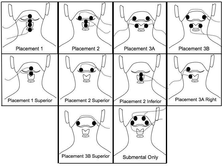
Figure 1.

shows the electrode positions relative to the hyoid bone, thyroid cartilage, and cricoid cartilage. The bipolar electrode pairs for each placement are connected by lead wires (dotted lines) with current flowing between the two electrodes of each pair. Placement 1, 2, 3A, and 3B have electrodes on both submental and laryngeal regions. Placements 1 superior, 2 superior, 2 inferior, 3A right, and 3B superior are individual electrode pairs. The submental-only placement has two electrode pairs above the hyoid bone on the submental region.
