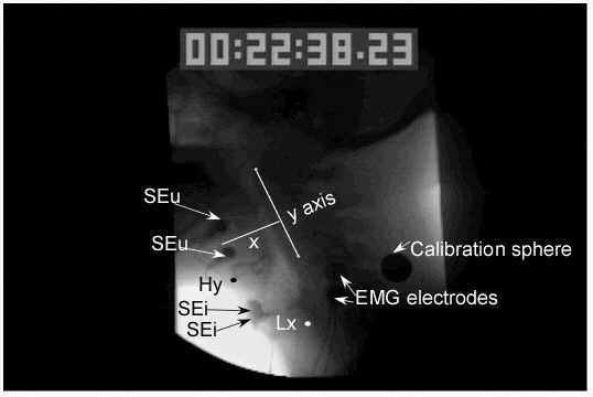
Figure 2.

shows the placement of the measurement points including the anterior inferior point on the hyoid bone (Hy) designated by a black dot and the posterior uppermost point of the subglottal air column to indicate the position of the larynx (Lx) designated by a white dot. Also shown is the y axis designated by a straight line drawn from the anterior inferior point of the first cervical vertebra to the anterior inferior point of the third vertebra. The x axis (x) was a straight line perpendicular to the y axis. A calibration sphere was taped to the side of the neck and surface electromyographic electrodes (EMG electrodes) were taped to the side of the neck and the stimulation artifact between them was used to determine when stimulation was turned on. The position 3B with two upper electrodes (SEu) and two inferior electrodes placed in the region of the thyroid cartilage (SEi) is shown.
