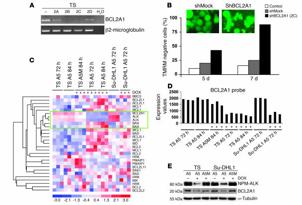
Figure 4. BCL2A1 expression is regulated by NPM-ALK activity and sustains the survival of ALK-positive ALCL cells.
(A and B) BCL2A1 shRNA induces apoptosis of ALCL cells. (A) TS cells were transduced with lentivirus (pLKO-GFP) expressing 4 BCL2A1-directed shRNAs (2A–D). BCL2A1 mRNA expression was determined by RT-PCR analysis (72 hours). (B) TS cells were transduced with a mock or a BCL2A1-directed shRNA (2C), and the percentage of apoptotic cells was determined by tetrametylrodamine methyl ester (TMRM) staining 5 and 7 days after infection. Cell morphology of BCL2A1- and mock shRNA–transduced TS cells is shown in the inset. These findings are representative of 3 independent experiments. (C–E) BCL2A1 expression correlates with ALK signaling. (C) The expression of BCL2 family genes was evaluated in all experimental conditions (33 samples) for absolute correlation with ALK. The BCL2A1 gene clustered within the branch including the ALK probe set (green rectangle). (D) BCL2A1 mRNA expression is significantly downmodulated after ALK RNAi. mRNA expression levels were assessed by microarray analysis of ALK-knockdown experiments in TS and Su-DHL1 cells. (E) Loss of ALK expression leads to the BCL2A1 protein downregulation. TS-TTA and Su-DHL1-TTA cells transduced with A5 or A5M were treated with DOX (84 hours). Western blot analysis was carried out on whole-cell lysates with antibodies against BCL2A1 and ALK.

