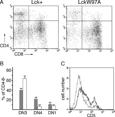
FIG. 4.
Analysis of early thymocyte development in lck+ and lckW97A mice. (A) Thymocytes were stained with antibodies against CD4, CD8, CD25, and CD44 and analyzed by flow cytometry. Dot plots show CD25 and CD44 staining of CD4− CD8− thymocytes. (B) Representation of DN3 (CD25+ CD44lo), DN4 (CD25− CD44−), and DN1 (CD25− CD44+) thymocytes in the CD4− CD8− population. Data are based on a quantitative analysis of 4 different pairs of lck+ and lckW97A mice. (C) Thymocytes were stained with antibodies against CD4, CD8, and CD5. Histograms show CD5 expression in CD4− CD8− population from lck+ (solid line) and lckW97A (dotted line) thymocytes. Data are representative of results from four separate pairs of animals.

