FIG. 1.
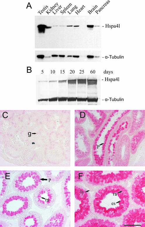
Expression profile and cellular localization of Hspa4l during testis development. (A and B) Immunoblot analysis of Hspa4l in cellular extracts from different tissues (A) and from testes of 5-, 10-, 15-, 20-, 25-, and 60-day-old mice (B), using polyclonal antibodies against mouse Hspa4l. A monoclonal antibody against α-tubulin was used as a loading control. (C to F) Immunohistochemistry using the Hspa4l antibody on sections of 5 (C)-, 15 (D)-, 25 (E)-, and 60-day-old (F) testes. Weak expression of Hspa4l was seen in gonocytes (g) of the 5-day-old testis (C). Hspa4l was highly expressed in pachytene spermatocytes (p) of the 15-day-old testis (D). In testes of 25- and 60-day-old mice (E and F), the protein was highly accumulated in spermatogenic cells, from late pachytene spermatocytes to postmeiotic round (rs) and elongated (es) spermatids. Hspa4l-immunopositive staining was barely detectable in Leydig (ly) cells and spermatogonia (sg). Bar, 100 μm.

