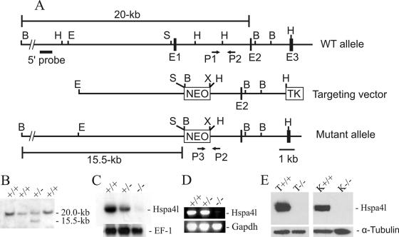
FIG. 2.
Targeted disruption of Hspa4l gene. (A) Structures of the wild-type, targeted-vector, and recombinant alleles are shown together with the relevant restriction sites. The numbers under the rectangles indicate the exons of Hspa4l. A 2.5-kb SmaI/HindIII fragment containing exon 1 was replaced by a pgk-neo selection cassette (NEO). The 5′ external probe used and the predicted length of BamHI restriction fragments in Southern blot analysis are shown. The primers P1, P2, and P3 used to amplify the wild-type and mutant alleles by PCR are also indicated. Abbreviations: TK, thymidine kinase cassette; B, BamHI; E, EcoRI; H, HindIII; S, SmaI; X, XhoI. (B) Southern blot analysis of recombinant ES cell clones. Genomic DNAs extracted from ES cell clones were digested with BamHI and probed with the 5′ probe shown in panel A. The Hspa4l wild-type allele generated a 20.0-kb BamHI fragment, whereas the targeted allele yielded a 15.5-kb BamHI fragment, as indicated in panel A. (C) Northern blot analysis. Total RNAs of Hspa4l−/−, Hspa4l+/−, and Hspa4l+/+ mice were hybridized with a cDNA probe containing the sequence of the 3′-untranslated region of Hspa4l. Rehybridization of blots with human elongation factor 1 cDNA (EF-1) revealed the integrity of RNA loading. (D) RT-PCR analysis using testicular RNA and primers located in exons 1 and 4 confirmed the absence of exon 1 in Hspa4l targeted transcripts. (E) Western blot with proteins extracted from testes (T) and kidneys (K) of Hspa4l+/+ and Hspa4l−/− mice, probed with anti-Hspa4l antibodies. The immunoreactive 94-kDa Hspa4l protein was detectable in wild-type but not in Hspa4l−/− tissues.

