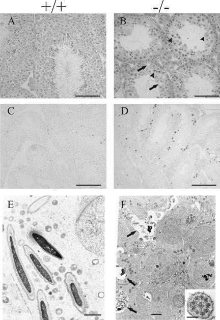
FIG. 4.
Spermatogenesis in the testes of Hspa4l−/− mice. Histological sections from testes of 16-week-old wild-type (A, C, and E) and infertile Hspa4l−/− (B, D, and F) mice are shown. (A and B) Staining with hematoxylin and eosin revealed hypospermatogenesis with a strongly reduced number of elongated spermatids in the seminiferous tubules of Hspa4−/− mice (B) compared to those of their wild-type littermates (A). Degenerated spermatogenic cells (arrowheads), vacuoles (arrows), a decreased diameter of tubules, and the presence of multinucleated giant cells (not shown) were observed in testes of infertile Hspa4l−/− mice (B). In situ TUNEL staining in the seminiferous tubules of wild-type (C) and Hspa4l−/− (D) males revealed an increase in adluminal cells with darkly stained nuclei in a subset of the tubules in mutant testes (D), while apoptotic cells in wild-type testes were most frequently located close to the basement membranes of seminiferous tubules (C). Electron micrographs document normal spermatogenesis with round and elongating spermatids in Hspa4l+/+ testes (E) and abnormal spermatogenesis in Hspa4l−/− testes (F). Arrows indicate several elongating spermatids at various stages of degeneration and vacuoles with cellular debris. Round spermatids reveal chromatin abnormalities and intracellular vacuoles, but these are much less prominent than in the later elongating spermatids. (Inset in panel F) Higher magnification of a cross section through a sperm flagellum showing the normal structure of the distal axoneme and microtubules. Bars: panels A and B, 200 μm; panels C and D, 100 μm; panel E, 2 μm; panel F, 1 μm; inset in panel F, 50 nm.

