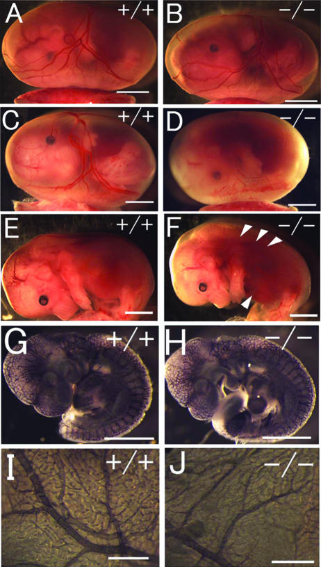
FIG. 2.
Gross morphology of Phd2−/− embryos. (A and B) E12.5 Phd2+/+ and Phd2−/− embryos with yolk sac. (C and D) E13.5 Phd2+/+ and Phd2−/− embryos with yolk sac. (E and F) E13.5 Phd2+/+ and Phd2−/− embryos after removal of yolk sacs. Phd2−/− embryos were alive at E12.5 and showed normal yolk sac vasculature. At E13.5, over 70% of Phd2−/− embryos were already dead, with a pale yolk sac. Arrowheads indicate dark-redness. (G to J) Whole-mount PECAM-1 immunohistochemistry. E9.5 Phd2+/− and Phd2−/− embryos (G and H). Images were captured by yolk sacs from E12.5 Phd2+/− and Phd2−/− embryos (I and J). Scale bars, 2 mm (A to F), 500 μm (G and H), 2 mm (I and J).

