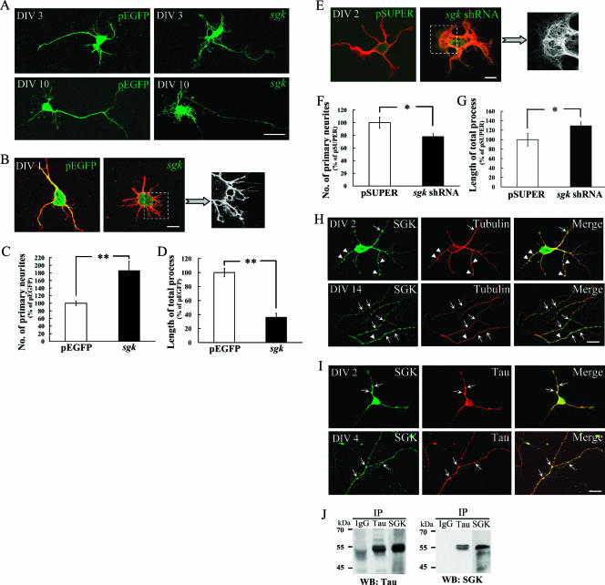
FIG. 1.
SGK1 increases neurite formation of hippocampal neurons. (A) Cultured hippocampal neurons were transfected with pEGFP-sgk1 at DIV 2 and DIV 9 and were fixed at DIV 3 and DIV 10, respectively. The EGFP-SGK1 protein expression was directly observed with a confocal fluorescence microscope. Scale bar, 20 μm. (B) Hippocampal neurons were transfected with pEGFP-sgk1 at DIV 0 and fixed at DIV 1. Neuronal processes were visualized by immunostaining with anti-β III tubulin Ab (red). The EGFP-SGK1 protein expression was readily observed with a confocal fluorescence microscope. Scale bar, 10 μm. (C and D) The transfection of pEGFP-sgk1 at DIV 0 significantly increased the number of primary neurites but decreased the length of the total process. n = 44 and 21 for pEGFP and pEGFP-sgk1 groups, respectively. Error bars indicate standard errors of the means. **, P was <0.01 compared with that of the control vector group. (E) Hippocampal neurons were transfected with pSUPER or pSUPER-sgk1 shRNA at DIV 0 and were fixed at DIV 2. Neuronal processes were visualized by immunostaining with anti-β III tubulin Ab (red). Scale bar, 10 μm. (F and G) The transfection of sgk1 shRNA decreased the number of primary neurites but increased the length of the total process. n = 10 and 19 for pSUPER and pSUPER-sgk1 shRNA groups, respectively. Error bars indicate standard errors of the mean. *, P was <0.05 compared with that of the control vector group. (H) Cultured hippocampal neurons were coimmunostained with SGK1 and tubulin Abs for confocal microscopic imaging. Endogenous SGK1 (green) is not only associated with MT (red) (arrowheads and yellow color) but also concentrated at regions devoid of stabilized MT (arrows and green color). The upper panels show DIV 2, and the lower panels show DIV 14. Scale bar, 20 μm. (I) SGK1 (green) is highly associated with tau (red) (arrows and yellow color). The upper panels show DIV 2, and the lower panels show DIV 4. Scale bar, 20 μm. (J) Endogenous tau and SGK1 were coimmunoprecipitated with each other. Anti-tau-protein G-Sepharose cross-linked Ab and anti-SGK1-protein G-Sepharose cross-linked Ab were incubated with 1,500 μg rat hippocampal lysate and subjected to 8% SDS-PAGE. PVDF membrane was probed with Abs specific for SGK1 and tau. Data are expressed as means ± standard errors of the means. Statistics were determined by Student's t test. IP, immunoprecipitate; WB, Western blot. IgG, immunoglobulin G.

