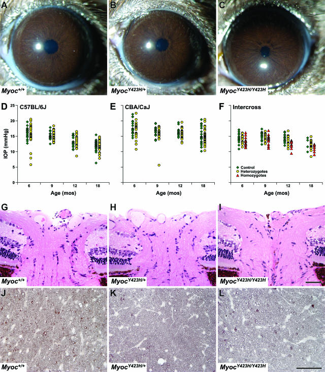
FIG. 2.
Assessment of control and mutant mice. (A to C) Anterior segments of all Myoc+/+, MyocY423H/+, and MyocY423H/Y423H mice were indistinguishable by slit lamp analysis regardless of genetic background (shown are samples from the C57BL/6J cross). (D to F) IOPs of MyocY423H/+ mice were indistinguishable from those of control mice at all ages tested regardless of genetic background. The numbers of control and mutant mice, respectively, at each age are as follows: for C57BL/6J mice, 20 and 44 at 6 months, 18 and 47 at 9 months; 17 and 29 at 12 months, and 26 and 34 at 18 months; and for CBA/CaJ mice, 20 and 27 at 6 months, 19 and 21 at 9 months, 14 and 29 at 12 months, and 23 and 43 at 18 months. Interestingly, MyocY423H/Y423H mice generated from intercrossing MyocY423H/+ mice from the C57BL/6J cross had IOPs that were slightly but significantly and consistently lower than those of controls at 6, 9, and 12 months (P = 0.030, P = 0.023, and P = 0.001, respectively). The absence of statistical significance at 18 months could be due to the relatively small number of control mice measured at this age, but there was a trend for MyocY423H/Y423H mice to be lower at this age as well. Numbers of Myoc+/+, MyocY423H/+, and MyocY423H/Y423H mice, respectively, for intercrosses are as follows: 16, 15, and 16 at 6 months; 14, 13, and 16 at 9 months; 13, 14, and 12 at 12 months; and 4, 18, and 8 at 18 months. Histological analysis did not reveal optic nerve head cupping (G to I) or retinal ganglion cell axon loss (J to L) in optic nerve cross-sections in any control or mutant mice analyzed. For eye histology, n = 3 for each genotype. For optic nerve cross-sections, n = 15 for Myoc+/+, n = 29 for MyocY423H/+, and n = 13 for MyocY423H/Y423H.

