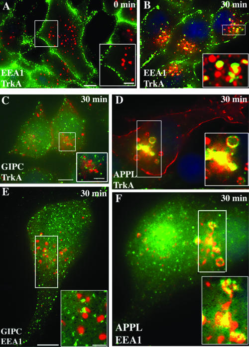
FIG. 5.
APPL but not GIPC traffics with TrkA to juxtanuclear, EEA1 endosomes. (A) After activation of TrkA and incubation at 4°C (0 min), TrkA (green) and EEA1 (red) staining do not overlap. (B) After 30 min at 37°C, TrkA (green) colocalizes with EEA1 (red) in juxtanuclear endosomes (yellow in inset). (C) At 30 min, GIPC (green) is distributed in a punctate pattern throughout the cytoplasm, and GIPC does not colocalize with TrkA in endosomes in the juxtanuclear region. (D) By contrast, colocalization of TrkA and APPL in juxtanuclear endosomes is striking (yellow in boxed area). (E and F) After 30 min, there is also a strong overlap between APPL (green) and EEA1 (red) in early endosomes (yellow pixels in boxed area in panel F) but little or no overlap between GIPC (green) and EEA1 in these endosomes (boxed area in panel E). Serum-starved PC12(615) cells were incubated at 4°C with NGF (E and F), anti-TrkA MAb 5C3 (C and D), or anti-TrkA RTA (A and B) and fixed (A) or shifted to 37°C for 30 min and then fixed (B to F). Cells were processed for immunofluorescence as described in the legend for Fig. 2. Incubation was done with rabbit anti-GIPC or anti-APPL IgG and mouse anti-EEA1 IgG, followed by appropriate secondary antibodies. Boxed regions in each panel are enlarged to the right. Bar = 2.5 μm; inset bar = 1 μm.

