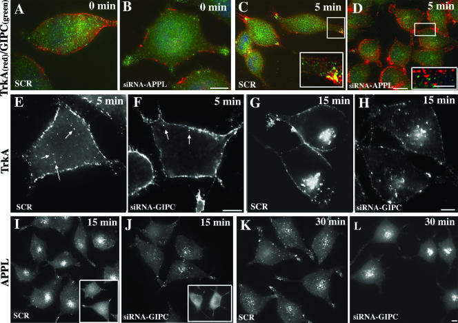
FIG. 6.
APPL and GIPC knockdown inhibits recruitment and trafficking of TrkA receptor. (A to D) APPL knockdown inhibits the recruitment of GIPC to TrkA endocytic vesicles. At 0 min after TrkA activation, TrkA does not colocalize with GIPC in PC12(615) cells transfected with either scrambled siRNA (A) or APPL siRNA (B). (C) Five minutes after TrkA activation, GIPC translocates to the cell membrane and colocalizes with TrkA vesicles in cells transfected with scrambled siRNA (SCR) but not in cells transfected with APPL siRNA (D). (E to H) GIPC knockdown slows down TrkA trafficking from the cell periphery to early endosomes in the juxtanuclear region. In control cells (SCR), at 5 min after TrkA activation, endocytic vesicles containing TrkA appear inside the cell (arrows in panel E), whereas in cells in which GIPC is knocked down (siRNA GIPC), the vesicles are still localized at the cell periphery (arrows in panel F). At 15 min, TrkA is concentrated in vesicles in the juxtanuclear region in controls (SCR) (G), but after GIPC knockdown (siRNA GIPC) there is less accumulation of TrkA vesicles in the juxtanuclear region (H). (I to L) GIPC knockdown slows down APPL trafficking. At 0 min after TrkA activation, APPL staining in both controls (SCR) and cells in which GIPC has been depleted (siRNA GIPC) is mostly cytoplasmic and diffuse (insets in panels I and J). At 15 min, APPL accumulates in vesicles in the juxtanuclear region in controls (SCR) (I) but not in cells in which GIPC has been knocked down (J). At 30 min, APPL staining in the juxtanuclear region is reduced in controls (SCR) (K) whereas in cells in which GIPC has been depleted (siRNA GIPC) APPL is concentrated on juxtanuclear vesicles (L). GIPC or APPL was depleted from PC12(615) cells by using siRNA as described in Materials and Methods, serum starved, and incubated at 4°C with an anti-TrkA antibody (5C3) to stimulate TrkA. They were then either fixed immediately (0 min) or shifted to 37°C for 5, 15, or 30 min and processed for immunofluorescence. Boxed regions in the panels are enlarged to the right. Bars, 2.5 μm (B, D, F, H, and L) and 1 μm (D, inset)

