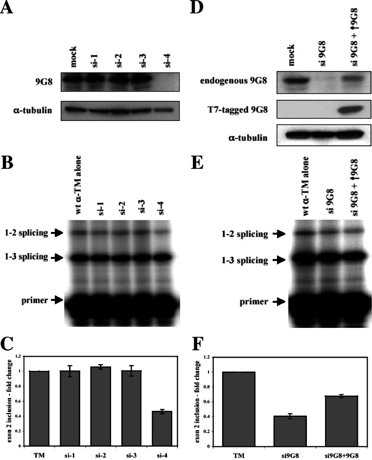
FIG. 7.
siRNA depletion of 9G8. (A) Wild-type pSVpA α-TM 1-4 minigene (400 ng) was cotransfected into HeLa cells with 20 nM concentrations of each of four individual siRNAs (si-1, si-2. si-3, and si-4) directed against 9G8. Western blots were performed on normal cells (mock) or cells treated with siRNAs against 9G8 using antibodies to 9G8 or α-tubulin. (B) Splicing patterns were analyzed as in Fig. 1, and the average and standard errors from at least three independent transfections are shown below (C). (D) Wild-type pSVpA α-TM 1-4 minigene (400 ng) was transfected into HeLa cells with either 20 nM of the si-4 siRNA directed against 9G8 or the si-4 siRNA plus cotransfection of an epitope-tagged (T7) version of 9G8. Western blots were performed on normal cells (mock) or cells treated with siRNAs against 9G8 using antibodies to 9G8, Τ7, or α-tubulin. (E) Splicing patterns were analyzed as in Fig. 1, and the average and standard errors from at least three independent transfections are shown at bottom (F).

