FIG. 1.
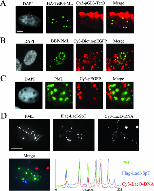
Plasmid DNA can be targeted to PML NBs. (A) Immunofluorescence micrographs of SK-N-SH cells transfected with Cy3-labeled pGL3-TetO and TetR-PMLIV constructs and detected with antibodies directed against an N-terminal HA tag. Chromatin was counterstained with DAPI. PML was detected by immunofluorescence. (B) SK-N-SH cells transfected with BBP-PMLIV and Cy3- and biotin-labeled pEGFP. Transfected cells were identified by GFP expression. (C) SK-N-SH cells transfected with BBP-PMLIV and Cy3-labeled, but not biotinylated, p-EGFP. (D) SK-N-SH cells transfected with Flag-LacI-SpT and Cy3-labeled plasmid DNA containing the LacO sequence. A line scan through several PML bodies (from left to right) reveals the relative distributions of PML protein, SpT protein, and the targeted DNA. Bars, 5 μm in panels A to C and 10 μm in panel D.

