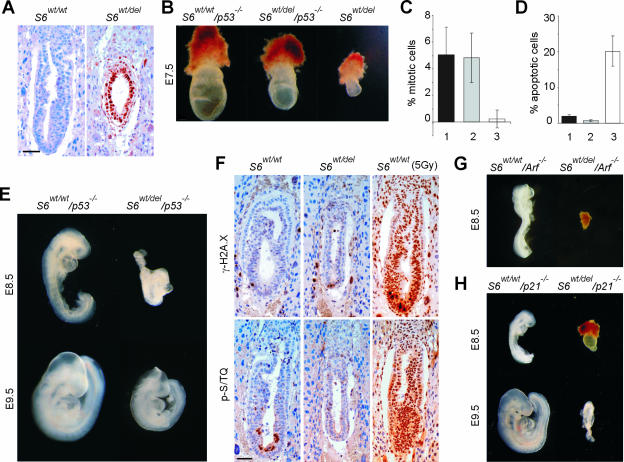
FIG. 3.
A p53-dependent checkpoint is activated in S6-heterozygous embryos. (A) Immunohistochemistry of sections of E6.5 S6wt/wt and S6wt/del embryos with antibodies against p53. (B) Morphology of representative E7.5 S6wt/wt/p53−/−, S6wt/del/p53−/−, and S6wt/del embryos. (C) Percentage of mitotic cells in the embryonic region of E7.5 S6wt/wt/p53−/− (n = 8), S6wt/del/p53−/− (n = 8), and S6wt/del (n = 7) embryos. (D) Percentage of apoptotic cells in E7.5 S6wt/wt/p53−/− (n = 8), S6wt/del/p53−/− (n = 7), and S6wt/del (n = 8) embryos. S6wt/wt/p53−/−, S6wt/del/p53−/−, and S6wt/del genotypes in C and D are indicated with 1, 2, and 3, respectively. (E) Whole-mount preparations of representative E8.5 and E9.5 S6wt/wt/p53−/− and S6wt/del/p53−/− embryos. (F) Immunohistochemistry of sections of E6.5 S6wt/wt and S6wt/del embryos with antibodies against the ATM/ATR phosphorylated consensus sequence (p-S/TQ) and phosphohistone H2A.X (γ-H2A.X). The majority of cells from E6.5 S6wt/wt embryos stained strongly positive with these antibodies 30 min after gamma irradiation (5Gy). (G) Morphology of representative S6wt/wt/p19Arf −/− (n = 14) and S6wt/del/p19Arf −/− (n = 11) embryos at E8.5. (H) Morphology of representative S6wt/wt/p21−/− and S6wt/del/p21−/− embryos at E8.5 and E9.5. Scale bar, 50 μm (A and F). Error bars in panels C and D denote SD. Embryos in panels B, E, G, and H were genotyped by PCR analysis.

