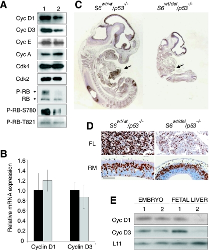
FIG. 6.
Characterization of the cell cycle progression in S6wt/del/p53−/− embryos. (A) Western blot analysis of lysates prepared from E10.5 S6wt/wt/p53−/− and S6wt/del/p53−/− embryos using antibodies against the indicated cell cycle regulators. Cyc, cyclin. (B) Quantitative RT-PCR analysis of cyclin D1 and cyclin D3 mRNAs expression in E10.5 S6wt/wt/p53−/− (black bars) and S6wt/del/p53−/− (gray bars) embryos. (C) Sagittal sections of the entire E11.5 embryos of indicated genotypes were stained with anti-BrdU antibody. Fetal livers are indicated by arrows. (D) BrdU incorporation in roofs of midbrain (RM) and fetal livers (FL) of the indicated genotypes (higher magnification of embryo sections from panel E). Scale bar, 100 μm. (E) Lysates of E12.5 fetal livers or the remaining parts of the embryo were analyzed by Western blotting employing antibodies against the indicated proteins. S6wt/wt/p53−/− and S6wt/del/p53−/− genotypes in panels A and E are indicated with 1 and 2, respectively.

