FIG. 1.
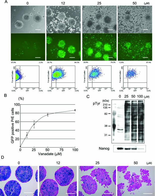
Increases in tyrosine phosphorylation induced primitive endoderm differentiation. (A) Afp-GFP ES cells were treated with sodium vanadate at the indicated concentrations for 48 h in the presence of LIF. Phase-contrast images are shown in the upper panels, and corresponding live fluorescence images are shown in the middle panels. Bars, 200 μm. The ES cells were dissociated with trypsin and analyzed by flow cytometry, which is shown in the lower panels. (B) Representative dose-response curve of sodium vanadate on primitive endoderm (PrE) differentiation. Primitive endoderm cells were estimated based on GFP expression in Afp-GFP ES cells by flow cytometry analysis. (C) Western blotting against antiphosphotyrosine (pTyr) antibody and anti-Nanog antibody. Protein tyrosine phosphorylation increased upon sodium vanadate treatment in Afp-GFP ES cells, whereas Nanog protein decreased. (D) Downregulation of Nanog expression in ES cells upon sodium vanadate treatment. Nanog β-geo ES cells were treated with the indicated concentrations of sodium vanadate for 48 h, and whole-mount X-Gal staining (blue) was performed. The paraffin sections were counterstained with eosin Y (pink). Bars, 100 μm.

