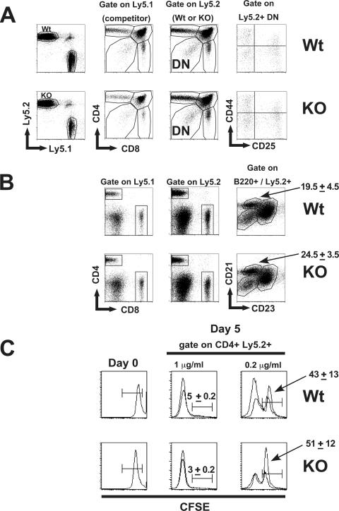
FIG. 3.
Normal T- and B-cell development in Dx1-KO stem cells. (A) Bone marrow pooled from pairs of Dx1-KO mice or littermate controls (both Ly5.2+) was mixed at a 1:1 ratio with bone marrow from normal (Ly5.1+) mice and used to reconstitute lethally irradiated recipients. After 3 months, thymus (A) was analyzed for expression of CD4+ and CD8+ cells among Ly5.1+ versus Ly5.2+ cells or for expression of CD44 and CD25 within the Ly5.2+ CD4− CD8− (DN) fraction. (B) Total splenocytes were analyzed for normal T- or B-cell development by expression of CD4, CD8, or B220. Immature, follicular, or marginal-zone B cells were identified within the B220+ fraction by expression of CD21 and CD23. Numbers are the average frequencies of marginal-zone cells found within the indicated region (two recipients per group). (C) In vitro proliferation of splenocytes from chimeric mice. Total splenocytes were labeled with CFSE and activated with plate-bound anti-CD3 at 1 μg/ml or 0.2 μg/ml. The level of CFSE fluorescence is shown for total splenocytes on day 0, or in the CD4+ Ly5.2+ fraction after 5 days of culture. Histograms show overlays for two mice analyzed per group, and numbers are the average values for the two mice analyzed.

